Vertebra Anatomy: Structure, Regional Variations, and Function
The vertebra is an irregular bone that serves as the fundamental building block of the vertebral column, commonly known as the spine. Found in all vertebrates, these complex structures are composed of bone and hyaline cartilage—a smooth, translucent cartilage found on the surfaces of joints. While the basic design is consistent across species, the proportions and specific shapes of vertebrae vary significantly depending on their position in the spine and the biological needs of the animal.
The primary role of the vertebral column is to provide a sturdy yet flexible axis for the body, protecting the delicate spinal cord while supporting the weight of the upper body.
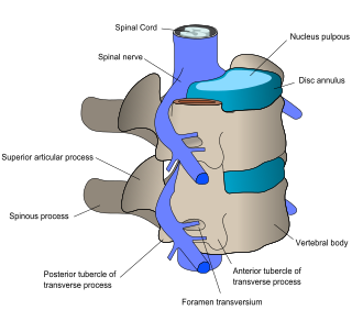
The Basic Structure of a Vertebra
A typical vertebra consists of two primary components: the vertebral body and the vertebral arch.
The Vertebral Body (Centrum)
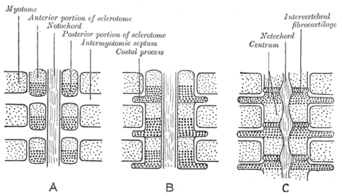
The vertebral body, or centrum, is the thick, anterior portion of the bone that bears the primary load of the spinal column. Its upper and lower surfaces are flattened and rough, providing the necessary attachment points for intervertebral discs, which act as shock absorbers between the bones.
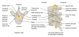
The Vertebral Arch
Located posterior to the body, the vertebral arch is a complex structure consisting of two pedicles and two laminae. Together with the body, the arch forms the vertebral foramen, a large central opening that creates the spinal canal to enclose and protect the spinal cord.
The arch also features seven projecting processes that facilitate movement and muscle attachment:
- One spinous process: Projects backward from the junction of the laminae.
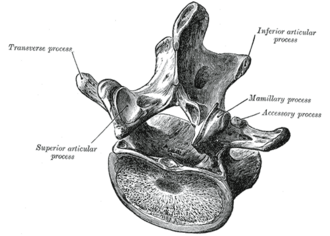
- Two transverse processes: Project laterally from the point where the lamina joins the pedicle.
- Four articular processes: Two superior and two inferior facets that restrict and guide the range of motion between adjacent vertebrae.
When vertebrae articulate, the notches formed by the pedicles create intervertebral foramina, which serve as the exit and entry conduits for spinal nerves.
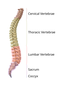
Regional Variations in the Human Spine
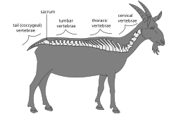
The human vertebral column typically consists of 33 vertebrae, though this number can range from 32 to 35 in about 10% of the population. As the vertebrae progress downward, they generally increase in size to accommodate greater weight-bearing loads, before becoming smaller again at the base.
Cervical Vertebrae (C1–C7)
Located in the neck, these seven vertebrae are small and delicate. They are characterized by transverse foramina—unique openings that allow vertebral arteries and veins to pass through toward the brain. The first two are highly specialized: C1 (the atlas) and C2 (the axis), which together enable the head to nod and rotate.
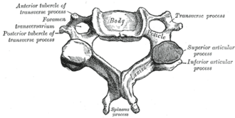
Thoracic Vertebrae (T1–T12)
The twelve thoracic vertebrae are heart-shaped and articulate with the ribs via specific costal facets. Because they are connected to the rib cage, their range of motion is more limited than that of the cervical or lumbar regions, primarily allowing for slight rotation.
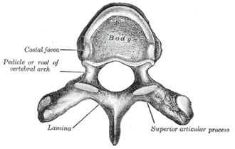
Lumbar Vertebrae (L1–L5)
The five lumbar vertebrae are the largest and strongest of the mobile vertebrae, designed to support the majority of the body's weight. Their transverse processes are sometimes called costiform processes because they correspond to rudimentary ribs not found in the thoracic region.
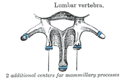
Sacral and Coccygeal Vertebrae
The base of the spine consists of five fused sacral vertebrae that form the sacrum, which connects the spine to the pelvis via the sacroiliac joints. Below the sacrum are three to five fused coccygeal vertebrae that form the coccyx, or tailbone.
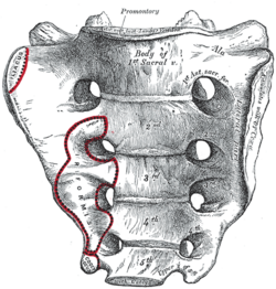
Key Facts
- Total Count: Usually 33 vertebrae in humans, divided into cervical (7), thoracic (12), lumbar (5), sacral (5 fused), and coccygeal (4 fused).
- Protection: The vertebral foramen protects the spinal cord, while intervertebral foramina allow spinal nerves to exit.
- Weight Bearing: The vertebral body (centrum) is the primary load-bearing part of the bone.
- Unique Features: Only cervical vertebrae possess transverse foramina for vertebral arteries.
- Mammalian Constant: Almost all mammals (except manatees and sloths) have exactly seven cervical vertebrae, regardless of neck length.
Comparative Anatomy in Other Animals
While the basic structure is similar, vertebrae vary across species. For example, birds typically have more cervical vertebrae (13–25) for increased neck flexibility. In contrast, whales often have fused cervical vertebrae to provide stability during swimming.
In non-mammalian vertebrates, such as lizards and some dinosaurs, cervical ribs are common. In mammals, the transverse processes are considered homologous to these cervical ribs.
Summary of Vertebral Regions
| Region | Number | Primary Characteristic | Key Function |
|---|---|---|---|
| Cervical | 7 | Transverse foramina; small size | Head support and mobility |
| Thoracic | 12 | Heart-shaped body; rib facets | Rib cage attachment; stability |
| Lumbar | 5 | Large, thick bodies | Weight bearing and support |
| Sacral | 5 (fused) | Fused into the sacrum | Pelvic connection |
| Coccygeal | 3–5 (fused) | Small, fused tailbone | Vestigial support |
Frequently Asked Questions
What is the difference between the atlas and the axis?
The atlas (C1) is the first cervical vertebra and supports the skull, allowing for the "yes" nodding motion. The axis (C2) is the second cervical vertebra and provides a pivot point that allows the head to rotate left and right.
What is spondylolysis?
Spondylolysis is a clinical defect occurring in the pars interarticularis, a thin portion of the vertebral arch. It most commonly affects the L5 (lowest lumbar) vertebra.
Why do some people have a different number of vertebrae?
Anatomical variations occur in about 10% of people. Common deviations include having 11 or 13 thoracic vertebrae, or 4 or 6 lumbar vertebrae, though the total count usually remains between 32 and 35.
How do vertebrae protect the spinal cord?
The vertebral body and the vertebral arch combine to form a hollow circle called the vertebral foramen. When stacked, these foramina create a continuous bony tunnel known as the spinal canal, which shields the spinal cord from external injury.