Vertebra Anatomy and Function in the Human Spine
The vertebra is a complex, irregular bone that serves as the fundamental building block of the vertebral column, commonly known as the spine. Composed of bone and hyaline cartilage—a smooth, pearly-white cartilage found on joint surfaces—these structures provide the essential framework that supports the body and protects the central nervous system.
While the basic configuration of a vertebra is consistent across most vertebrate species, the proportions and specific shapes vary depending on the spinal segment and the animal's biological needs. In humans, these bones are meticulously organized to balance the competing needs of structural stability and flexible movement.
Key Facts
- Primary Function: Supports the body's weight and protects the spinal cord.
- Human Count: Typically 33 vertebrae, though this can range from 32 to 35.
- Core Components: Each vertebra consists of a load-bearing body (centrum) and a protective vertebral arch.
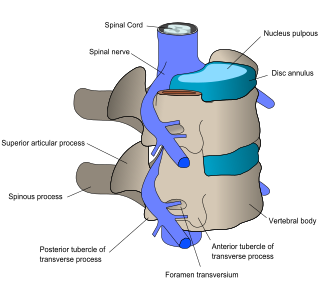
- Nerve Passages: Intervertebral foramina allow spinal nerves to exit the column.
- Regional Variation: Vertebrae increase in size as they move downward toward the lumbar region to handle increased weight.
The Basic Structure of a Vertebra
A typical vertebra is divided into two primary sections: the vertebral body and the vertebral arch.
The Vertebral Body (Centrum)
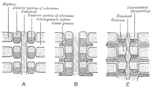
The vertebral body, or centrum, is the thick, anterior portion of the bone. Its primary role is to bear the weight of the body. The upper and lower surfaces of the body are rough and flattened, providing the necessary attachment points for intervertebral discs, which act as shock absorbers between the bones.
The Vertebral Arch
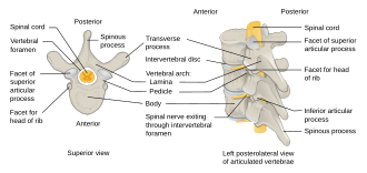
Located posterior to the body, the vertebral arch consists of two pedicles and two laminae. Together with the body, the arch forms the vertebral foramen, a large central opening that creates the spinal canal to enclose and protect the spinal cord.
The arch also features seven projecting processes that facilitate movement and muscle attachment:
- One Spinous Process: Projects backward and is often palpable under the skin.
- Two Transverse Processes: Project laterally from the sides.
- Four Articular Processes: These form facet joints (superior and inferior) that restrict and guide the range of spinal motion.
Regional Variations of the Human Spine
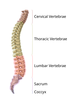
The human vertebral column is divided into five distinct regions, each adapted for specific mechanical roles.
Cervical Vertebrae (C1–C7)
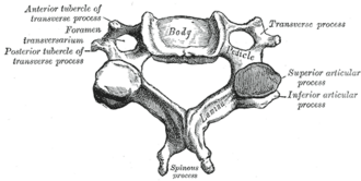
Located in the neck, these seven vertebrae are the smallest and lightest. They are characterized by transverse foramina—unique openings that allow vertebral arteries to pass through to the brain. The first two are highly specialized: C1 (the atlas) and C2 (the axis), which together enable the head to nod and rotate.
Thoracic Vertebrae (T1–T12)
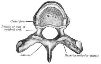
The twelve thoracic vertebrae are heart-shaped and articulate with the ribs via specific costal facets. Because they are attached to the rib cage, they offer significantly less flexibility than the cervical or lumbar regions, primarily allowing for slight rotation.
Lumbar Vertebrae (L1–L5)
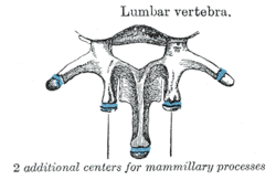
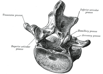
The five lumbar vertebrae are the largest and strongest, designed to support the majority of the body's weight. They feature prominent mammillary processes and lack the facets for rib attachment found in the thoracic region.
Sacral and Coccygeal Vertebrae
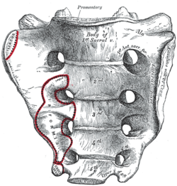
At the base of the spine, five sacral vertebrae fuse in adulthood to form the sacrum, which connects the spine to the pelvis via the sacroiliac joints. Below the sacrum are three to five fused coccygeal vertebrae, forming the coccyx, or tailbone.
Comparative Anatomy and Development
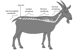
While humans have a standardized count of cervical vertebrae, this is a trait shared by almost all mammals (including giraffes and whales), except for manatees and sloths. In contrast, birds typically possess 13 to 25 cervical vertebrae to allow for extreme neck flexibility.
In non-mammalian vertebrates, such as lizards or certain dinosaurs, cervical ribs may be present, whereas in mammals, these have evolved into the transverse processes.
Summary of Vertebral Regions
| Region | Number | Key Characteristic | Primary Function |
|---|---|---|---|
| Cervical | 7 | Transverse foramina | Head support and mobility |
| Thoracic | 12 | Rib articulation facets | Protection of thoracic organs |
| Lumbar | 5 | Large, thick bodies | Weight bearing |
| Sacral | 5 (fused) | Forms the sacrum | Pelvic stability |
| Coccygeal | 3–5 (fused) | Forms the coccyx | Vestigial tail structure |
Frequently Asked Questions
What is the difference between the atlas and the axis?
The atlas (C1) is the first cervical vertebra and supports the skull, allowing for the "yes" nodding motion. The axis (C2) acts as a pivot for the atlas, enabling the "no" rotating motion of the head.
What is spondylolysis?
Spondylolysis is a clinical defect occurring in the pars interarticularis, a thin portion of the vertebral arch. It most commonly affects the L5 (lowest lumbar) vertebra.
Why do vertebrae increase in size as they go down the spine?
Vertebrae increase in size to accommodate the increasing load of body weight. The lumbar vertebrae must support the weight of the entire upper torso, requiring a larger, denser vertebral body.
What are intervertebral foramina?
Intervertebral foramina are the openings created when the notches of two adjacent vertebrae articulate. These serve as the exit and entry conduits for spinal nerves leaving the spinal cord.