Spinal Column: Anatomy, Structure, and Evolutionary Variations
The spinal column, also known as the vertebral column, is a complex skeletal structure found in approximately 50,000 species of animals. In humans, the spine serves as the central support for the body and a protective conduit for the spinal cord. While the general structure of human vertebrae is typical of most mammals, reptiles, and birds, the specific shape and number of vertebrae can vary significantly across different species and even between individuals.
The human spine is composed of a series of bones called vertebrae, which are organized into distinct regions. These bones provide the necessary rigidity to support the torso while maintaining the flexibility required for movement.
Key Facts
- Total Vertebrae: Normally 33 in humans (ranging from 32 to 35).
- Articulating Bones: The upper 24 pre-sacral vertebrae are separate and joined by intervertebral discs.
- Fused Bones: The lower nine vertebrae (sacrum and coccyx) are typically fused in adults.
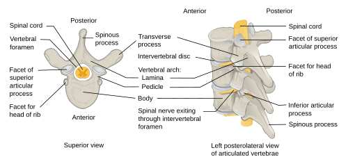
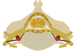
- Protective Function: The vertebral foramen aligns to form the spinal canal, which houses the spinal cord.
- Development: The spine forms during embryogenesis through a process called somitogenesis.
The Structure of the Human Spine
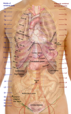
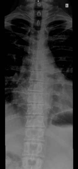
The human spinal column is divided into five primary regions, each with specific characteristics and functions. The articulating vertebrae in these regions are generally similar but vary slightly to accommodate the needs of the area.
Regional Breakdown
- Cervical Spine (Neck): Consists of 7 vertebrae (C1–C7). This region includes the atlas and axis, the two specialized vertebrae that support the head.
- Thoracic Spine (Chest/Upper Back): Consists of 12 vertebrae (T1–T12). These vertebrae feature facets for articulation with the ribs.
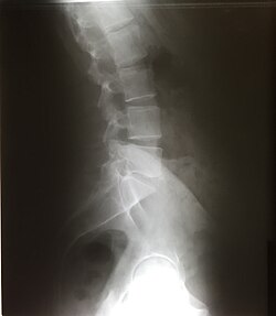
- Lumbar Spine (Lower Back): Consists of 5 vertebrae (L1–L5). These are designed to withstand higher axial structural loads.
- Sacrum (Pelvis): Consists of 5 fused vertebrae (S1–S5).
- Coccyx (Tailbone): Consists of 3 to 5 fused vertebrae.
Anatomy of a Single Vertebra
A typical vertebra is composed of two main parts: the vertebral body (centrum), located ventrally to handle structural loads, and the vertebral arch (neural arch), located dorsally to provide anchorages for muscles and ribs. Together, these form the vertebral foramen, which creates the spinal canal.
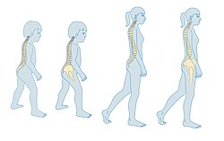
Because the spinal column grows faster than the spinal cord during childhood, the spinal cord typically ends at the upper lumbar spine (around the L1/L2 level) by adulthood. The remaining space in the canal is occupied by the cauda equina, a bundle of spinal nerves resembling a horse's tail.
Ligaments and Support
To maintain stability and facilitate movement, the spinal column is held together by several key ligaments:
- Longitudinal Ligaments: The anterior and posterior longitudinal ligaments run the length of the column along the front and back of the vertebral bodies.
- Ligamentum Flavum: Located deep to the laminae.
- Interspinous and Supraspinous Ligaments: These connect and run along the spinous processes.
- Intertransverse Ligaments: These connect the transverse processes.
Development and Embryogenesis
The segmented pattern of the spine is established during embryogenesis through somitogenesis. Around the third week of development, spheres of paraxial mesoderm called somites are added to the posterior of the embryo. In humans, there are typically 42 to 44 somites.
Sclerotomes (precursors to bone) migrate to surround the notochord and spinal cord. Through a process called resegmentation, the posterior part of one somite fuses with the anterior part of the next to form the definitive vertebrae. Disruptions in this process, often linked to mutations in genes such as MESP2, DLL3, and LFNG, can lead to congenital scoliosis.
Clinical Significance and Common Conditions
The spine is susceptible to various congenital and acquired conditions that can affect mobility and neurological function.
Curvature Abnormalities
Scoliosis is a lateral curvature of the spine affecting approximately 0.5% of the population. It is more common in females and can result from unequal vertebral growth or pulmonary atelectasis (partial lung collapse).
Structural Defects
- Spinal Stenosis: A narrowing of the spinal canal that can constrict the spinal cord and cause neurological deficits.
- Spondylolysis: A fracture or defect at the pars interarticularis of the vertebral arch.
- Klippel–Feil Syndrome: A rare congenital condition where two or more cervical vertebrae are fused.
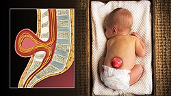
- Spina Bifida: A congenital defect where the vertebral arch fails to close.
Comparative Anatomy in Other Animals
While the basic blueprint of the vertebral column is conserved across vertebrates, significant variations exist based on evolutionary needs.
| Species/Group | Notable Characteristic | Example/Detail |
|---|---|---|
| Mammals | Consistent cervical count | Almost all have 7 cervical vertebrae (except sloths and manatees). |
| Birds | Variable cervical count | Swans have 25; some have a fused synsacrum. |
| Snakes | Extreme thoracic count | Can possess hundreds of thoracic vertebrae and ribs. |
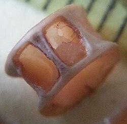
| Dinosaurs | Weight reduction | Pleurocoels (hollow depressions) in sauropods reduced weight by over a ton. |
| Plesiosaurs | Extreme neck length | Elasmosaurus had 76 cervical vertebrae. |
In dinosaurs, the presence of three or more sacral vertebrae is a defining characteristic. Some species also utilized ossified tendons to reinforce their caudal (tail) vertebrae.
Frequently Asked Questions
How many vertebrae are in the average human spine?
A typical human spinal column has 33 vertebrae, though this can range from 32 to 35 in about 10% of the population due to natural variations in the thoracic, lumbar, or coccygeal regions.
What is the difference between the atlas and the axis?
The atlas (C1) and axis (C2) are the two most superior vertebrae in the cervical spine; they are specialized to support the skull and allow for the rotation and tilting of the head.
What is the cauda equina?
The cauda equina is a bundle of spinal nerves at the lower end of the spinal canal. It forms because the vertebral column outgrows the spinal cord during development, leaving the cord to end around the L1/L2 level.
What causes congenital scoliosis?
Congenital scoliosis is often caused by disruptions during somitogenesis in the embryo, which can be linked to mutations in specific genes like MESP2, DLL3, and LFNG.
Why do some animals have more cervical vertebrae than humans?
While most mammals are limited to seven, other vertebrates evolved different counts to suit their environments; for example, swans have 25 and the extinct Elasmosaurus had 76 to support an extremely long neck.