Water Vascular System of Echinoderms: Mechanics and Anatomy
The water vascular system (also known as the hydrovascular, ambulacral, or aquiferous system) is a unique hydraulic network found in echinoderms, such as sea stars and sea urchins. This sophisticated system serves multiple critical biological functions, including locomotion, respiration, and the transportation of food and waste.
At its core, the system consists of a network of canals that connect to numerous tube feet. Echinoderms move by alternately contracting muscles to force water into these feet, causing them to extend and push against a surface. When the muscles relax, the feet retract, allowing the animal to pull itself forward. This process is powerful, though typically slow.
The water vascular system is integrated into the animal's coelomic cavities, working alongside the haemal, perivisceral, gonadal, and perihaemal coeloms. While the fluid within the system is primarily sea water, it also contains proteins, high levels of potassium salts, and coelomocytes—amoeboid cells similar to vertebrate blood cells.
Key Facts
- Primary Functions: Used for movement, breathing, and transporting nutrients and waste.
- Mechanism: Operates via hydraulic pressure created by muscle contractions in the ampullae.
- Key Components: Typically includes a madreporite, stone canal, ring canal, radial canals, and tube feet.
- Fluid Composition: Mostly sea water enriched with potassium salts, proteins, and coelomocytes.
- Diversity: Structure varies significantly across the five echinoderm classes.
Anatomy in Sea Stars
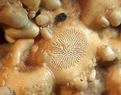
In sea stars, water enters through the madreporite, a sieve-like structure located on the animal's upper surface. This leads to a small sac called an ampulla, which connects to the stone canal—a duct often lined with calcareous material.
The stone canal feeds into a circular ring canal, which distributes water to radial canals extending along the ambulacral grooves on the underside of each arm. Branching off these radial canals are bulb-like ampullae connected via lateral canals. These ampullae lead to sucker-like podia; together, the ampulla and podium form a tube foot. In sea stars, these are usually staggered along the canal.
The central ring canal also contains specialized structures: polian vesicles (muscular sacs) and Tiedemann's bodies (complex pouches that produce coelomocytes).
Variations Across Other Echinoderms
Ophiuroids (Brittle Stars and Basket Stars)
Ophiuroids differ from sea stars in several ways. Their madreporite is located on the underside, usually within a jaw plate. They lack ambulacral grooves, and their radial canals run through solid bone-like ossicles in the arms. Additionally, they do not have ampullae; instead, they use a simple valve at the top of the foot to regulate water pressure.
Sea Urchins
The madreporite of a sea urchin is found on the upper surface within the plates surrounding the anus. The stone canal descends to a ring canal surrounding the oesophagus. Because sea urchins lack arms, their five radial canals run along the inside of the skeletal "test" (shell). Their tube feet penetrate the test in ten rows and are often highly modified for specific tasks.
Crinoids
Crinoids are unique because they lack a madreporite. Instead, they use numerous minute ciliated funnels on the oral surface to bring water into the body cavity. While they have small stone canals, these open internally. They lack ampullae, maintaining water pressure through the ring canal, which is surrounded by contractile muscle fibres.
Sea Cucumbers
In sea cucumbers, the system is closed off from the outside and is filled with internal coelomic fluid. The madreporite is located internally, just below the pharynx. While most have radial canals and ampullae leading to tube feet and oral tentacles, the order Apodida lacks both tube feet and radial canals, with tentacles branching directly from the ring canal.
Comparative Summary of Water Vascular Systems
| Class/Group | Madreporite Location | Ampullae Present? | Key Characteristic |
|---|---|---|---|
| Sea Stars | Upper surface | Yes | Staggered tube feet in ambulacral grooves |
| Ophiuroids | Underside (jaw plate) | No | Canals run through solid ossicles |
| Sea Urchins | Upper surface (near anus) | Yes | Tube feet penetrate the skeletal test |
| Crinoids | None (ciliated funnels) | No | Pressure maintained by ring canal muscles |
| Sea Cucumbers | Internal (below pharynx) | Yes | Filled with internal coelomic fluid |
Frequently Asked Questions
What is the primary purpose of the water vascular system?
It is a hydraulic system used by echinoderms for locomotion, respiration, and the transportation of food and waste products throughout the body.
How do tube feet actually move the animal?
Muscles contract to force water into the tube feet, causing them to extend and push against the ground. When the muscles relax, the feet retract, pulling the animal forward.
Do all echinoderms have a madreporite?
No. While most do, crinoids lack a madreporite and instead use ciliated funnels to move water into the body cavity.
What are coelomocytes?
Coelomocytes are amoeboid cells produced by Tiedemann's bodies in the ring canal; they are functionally similar to the blood cells found in vertebrates.
How does the system in sea cucumbers differ from sea stars?
Unlike sea stars, the water vascular system in sea cucumbers has no direct connection to the outside environment and is filled with internal coelomic fluid rather than sea water.