Serous Membrane
The serous membrane, also known as the serosa or tunica serosa, is a specialized smooth epithelial membrane that lines the inner walls of body cavities and covers the internal organs within them. Its primary purpose is to secrete a lubricating serous fluid, which allows organs to slide smoothly against opposing surfaces without friction.
Unlike the adventitia—a connective tissue layer used to bind structures together—the serous membrane is specifically designed to reduce friction. This is critical for organs that move constantly, such as the beating heart or the inflating lungs, as friction could otherwise lead to inflammation.
Key Facts
- Composition: Consists of a single layer of simple squamous epithelium (mesothelium) and an underlying connective tissue layer.
- Dual Layers: Divided into the parietal layer (lining the cavity wall) and the visceral layer (covering the organ).
- Function: Secretes serous fluid to provide lubrication and prevent friction between moving organs.
- Origin: Derived from the mesoderm of the trilaminar embryo.
- Major Cavities: Found in the pericardial, pleural, and peritoneal cavities.
Structure and Composition
Every serous membrane is composed of two distinct layers that work together to protect internal organs.
The Epithelial Layer (Mesothelium)
The surface layer is the mesothelium, which consists of a single layer of avascular flat nucleated cells known as simple squamous epithelium. These cells produce the lubricating serous fluid, which has a consistency similar to thin mucus.
The Connective Tissue Layer
Beneath the mesothelium lies a layer of connective tissue. This layer is essential because it provides the necessary blood vessels and nerves for the secretory cells and acts as the binding agent that allows the membrane to adhere to organs and body walls.
Visceral vs. Parietal Layers
Serous membranes are organized into two layers that create a potential space between them.
- Parietal Layer: This layer lines the walls of the body cavity.
- Visceral Layer: This layer wraps directly around the internal organs (the viscera).
The space between these two layers is called the serous cavity. This space is mostly empty, containing only a small amount of serous fluid to facilitate movement.
![Serous membrane lines the pericardial cavity and reflects back to cover the heart—much the same way that an underinflated balloon would form two layers surrounding a fist.[1]](/images/d8/3a/d83ac308913eacbffd1a1e2f71246e9452f0ee43df44b0d6f83d5e0ee6b2fece.jpg)
Major Serous Membranes in the Human Body
There are three primary serous cavities in the human body, each with its own specific nomenclature:
The Pericardium
The pericardium is the two-layered sac surrounding the heart. It consists of the parietal pericardium and the visceral pericardium (also called the epicardium). It surrounds the entire heart except where blood vessels emerge from the superior side.
The Pleura
The pleura lines the thoracic cavity and surrounds the lungs. It is divided into the parietal pleura and visceral pleura. In this cavity, the lubricating role of the membrane is especially vital for the mechanics of breathing.
The Peritoneum
The peritoneum lines the abdominopelvic cavity. The parietal peritoneum attaches to the abdominal and pelvic walls, while the visceral peritoneum wraps around the abdominal organs.
Other specialized examples include the perimetrium (the serosa of the uterus) and the tunica vaginalis (the serous membrane surrounding the testes).
Embryonic Development
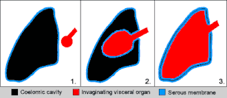
All serous membranes originate from the mesoderm of the trilaminar embryo. During development, the lateral plate mesoderm splits to form a cavity known as the intraembryonic coelom.
This split creates two distinct layers:
- Splanchnopleure: Associated with the endoderm, this layer becomes the visceral serous membrane.
- Somatopleure: Associated with the ectoderm, this layer becomes the parietal serous membrane.
As the embryo develops, organs invaginate into the bag-like coelom, ensuring they are surrounded by the serous membrane rather than sitting inside the cavity itself.
Summary of Serous Membranes
| Membrane Name | Cavity Location | Organ Covered | Specific Layers |
|---|---|---|---|
| Pericardium | Pericardial Cavity | Heart | Parietal & Visceral Pericardium |
| Pleura | Pleural Cavity | Lungs | Parietal & Visceral Pleura |
| Peritoneum | Abdominopelvic Cavity | Abdominal Organs | Parietal & Visceral Peritoneum |
| Perimetrium | Pelvic Region | Uterus | Visceral Serosa |
| Tunica Vaginalis | Scrotal Sac | Testis | Serous Membrane |
Clinical Significance
Certain diseases specifically target these membranes. Mesotheliomas are neoplasms (tumors) that occur in the serous membranes. Additionally, the modified Müllerian-derived serous membranes surrounding the ovaries can develop into serous tumors, which may be solid or papillary in nature and can also appear within the uterus.
Frequently Asked Questions
What is the difference between the visceral and parietal layers?
The parietal layer lines the wall of the body cavity, while the visceral layer covers the actual organ. The space between them contains serous fluid.
How does a serous membrane differ from adventitia?
A serous membrane secretes fluid to reduce friction between moving surfaces, whereas adventitia is a connective tissue layer used to bind structures together.
What is the role of the mesothelium?
The mesothelium is the simple squamous epithelial layer of the serous membrane responsible for producing the lubricating serous fluid.
Where do serous membranes come from during development?
They develop from the mesoderm of the trilaminar embryo, specifically from the splanchnopleure (visceral) and somatopleure (parietal) layers surrounding the intraembryonic coelom.
What happens if the serous membrane fails to lubricate an organ?
Without the lubricating serous fluid, the friction caused by the movement of organs (like the heart beating or lungs expanding) could lead to inflammation of the organs.