Mesothelium: Structure, Function, and Clinical Significance
The mesothelium is a specialized membrane composed of simple squamous epithelial cells. Originating from the mesoderm, this thin but vital layer forms the lining of several critical body cavities, ensuring that internal organs can move smoothly without friction. When the mesothelium functions primarily to secrete serous fluid, it is referred to as a serosa.
This membrane is strategically located in three primary areas: the pleura (surrounding the lungs), the pericardium (surrounding the heart), and the peritoneum (lining the abdominopelvic cavity, including the mesentery, falciform ligament, greater and lesser omentum, and perimetrium). Additionally, it surrounds the male testis as the tunica vaginalis and may occasionally be found in the spermatic cord within a patent processus vaginalis.
Anatomically, the mesothelium is categorized based on its location: visceral mesothelium covers the internal organs themselves, while parietal mesothelium lines the surrounding body walls.
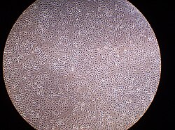
Key Facts
- Origin: Derived from the somatopleuric mesenchyme of the embryonic mesoderm.
- Structure: A monolayer of flattened squamous-like cells resting on a basement membrane.
- Primary Role: Secretes lubricating serous fluid to facilitate intracoelomic movement.
- Clinical Link: Over 90% of mesothelioma cases are linked to asbestos exposure.
- Immune Function: Mesothelial cells act as antigen-presenting cells and are capable of phagocytosis.
Origin and Biological Structure
The mesothelium develops from the embryonic mesoderm cell layer that lines the coelom (the embryonic body cavity). This developmental path allows it to create a protective barrier for the majority of the body's internal organs.
Structurally, the mesothelium consists of a single layer of flattened cells supported by a thin basement membrane and dense irregular connective tissue. While typically squamous, these cells may become cuboidal in specific areas, such as the peritoneal side of the diaphragm overlaying lymphatic lacunae, the milky spots of the omentum, or at sites of injury.
The luminal surface of these cells is covered in microvilli. These tiny projections trap proteins and serosal fluid, creating the slippery surface necessary for organs to slide past one another during movement.
Physiological Functions
The primary purpose of the mesothelium is the production of a lubricating fluid. This fluid is released between layers to create a non-adhesive, protective surface that prevents friction during the movement of organs within the body cavities.
Beyond lubrication, the mesothelium performs several complex biological roles:
- Transport: It facilitates the movement of fluid and particulate matter across serosal cavities.
- Immune Response: It enables leukocyte migration in response to inflammatory mediators and functions as an antigen-presenting cell.
- Repair: It synthesizes growth factors, pro-inflammatory cytokines, and extracellular matrix proteins to assist in serosal repair.
- Fibrin Clearance: It releases factors like plasminogen to promote the disposition and clearance of fibrin.
- Protection: The secretion of lubricants and glycosaminoglycans may help protect the body against tumor dissemination and infection.
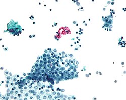
![Cytology of reactive mesothelium, with typical features. The main causes of reactive mesothelium are infection, trauma and cancer.[3] Wright's stain.](/images/2f/e3/2fe3978626197749234a7517917df7ce4ad7a49db9a31b12cb53fafe4d1e299d.jpg)
Clinical Significance and Disease
When the mesothelium is compromised or becomes abnormal, several serious medical conditions can arise.
Mesothelioma
Mesothelioma is a cancer where mesothelial cells divide uncontrollably, invading nearby tissues and organs. These malignant cells can metastasize to other parts of the body, most commonly originating in the peritoneum or pleura. Asbestos exposure is the primary risk factor, linked to more than 90% of cases.
Intra-abdominal Adhesions
Under normal conditions, the mesothelium secretes plasminogen to remove fibrin deposits. However, surgical trauma can damage the mesothelium, reducing its fibrinolytic capacity. This leads to the accumulation of fibrin and the formation of fibrous adhesions. In the abdomen, these can cause female infertility or intestinal obstruction; in the thorax, they may impair lung and heart function.
Ultrafiltration Failure
In patients undergoing peritoneal dialysis, the mesothelium can undergo fibrosis. This is often driven by acidity, supra-physiological glucose concentrations, and glucose degradation products in the dialysis fluid. This fibrosis occurs via the proliferation of fibroblasts or epithelial–mesenchymal transition, resulting in increased solute passage and the eventual failure of ultrafiltration.
| Feature | Description |
|---|---|
| Cell Type | Simple squamous epithelium (can become cuboidal during injury) |
| Embryonic Origin | Somatopleuric mesenchyme (Mesoderm) |
| Key Locations | Pleura, Peritoneum, Pericardium, Tunica vaginalis |
| Primary Secretion | Serous fluid and Plasminogen |
| Major Pathology | Mesothelioma (often asbestos-linked) |
Frequently Asked Questions
What is the difference between visceral and parietal mesothelium?
Visceral mesothelium is the layer that directly covers the internal organs, whereas parietal mesothelium lines the walls of the body cavities surrounding those organs.
How does asbestos lead to mesothelioma?
While the source does not detail the cellular mechanism, it notes that more than 90% of mesothelioma cases—a cancer where mesothelial cells divide without control—are linked to asbestos exposure.
What happens when the mesothelium cannot clear fibrin?
If the mesothelium is damaged (e.g., during surgery) and cannot secrete enough plasminogen to remove fibrin, fibrous adhesions can form. These adhesions may lead to intestinal obstruction, infertility, or impaired cardiac and pulmonary function.
Why do peritoneal dialysis patients experience ultrafiltration failure?
Failure occurs when the peritoneal mesothelium becomes fibrosed due to the acidity and high glucose levels in dialysis fluids, which increases the passage of solutes and reduces the efficiency of fluid removal.
What is the role of microvilli in the mesothelium?
Microvilli on the luminal surface trap proteins and serosal fluid, which creates the slippery, non-adhesive surface that allows internal organs to slide past each other without damage.