Heart Anatomy, Physiology, and Cardiovascular Health
The heart is the central engine of the circulatory system, a muscular organ responsible for pumping blood throughout the body to deliver oxygen and nutrients while removing waste. Given its critical role, the health of the heart is paramount to overall survival. In recent years, cardiovascular diseases have become the leading cause of death globally, making it essential to understand how this complex organ functions and how to maintain its health.

Key Facts
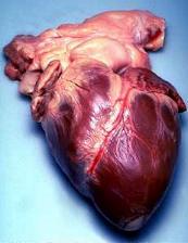
- Global Impact: Cardiovascular diseases caused an estimated 19.8 million deaths in 2022, accounting for roughly 32% of all global deaths.
- Physical Specs: An adult heart typically weighs between 250–350 grams and is roughly the size of a closed fist.
- Heart Rate: A normal adult resting heart rate ranges from 60 to 100 beats per minute (bpm).
- Primary Risks: Smoking, obesity, physical inactivity, high cholesterol, hypertension, and poorly controlled diabetes are major risk factors for heart disease.
- Medical Specialists: Doctors specializing in heart health are known as cardiologists.
Anatomy and Structure
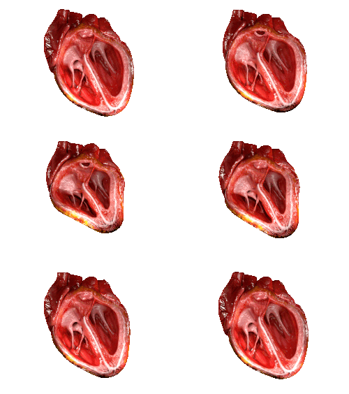
Location and Shape
The heart is a cone-shaped organ located in the middle of the thorax. Its base is positioned upward, tapering down to a point called the apex. While it is centrally located, the largest portion is usually offset to the left side of the chest, a condition known as levocardia. In rare cases, a congenital disorder called dextrocardia causes the heart to be offset to the right.
![The human heart is in the middle of the thorax, with its apex pointing to the left.[14]](/images/f3/19/f3190feff33bfb1c17c1fd773cdc92e6a25552de08cbfa21601e571c45836d27.png)
Because the heart occupies space between the lungs, the left lung is slightly smaller than the right and features a cardiac notch to accommodate the organ's position.
The Heart Wall and Pericardium
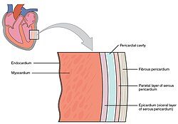
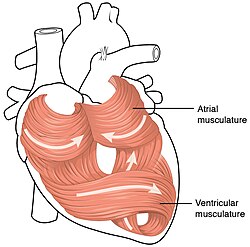
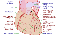
The heart is encased in a protective sac called the pericardium, which consists of visceral and parietal layers. The muscular wall of the heart is composed of the myocardium, which features a swirling pattern that allows the heart to pump blood with maximum efficiency.
Chambers and Valves
The human heart is divided into four chambers: two upper atria and two lower ventricles. The right side of the heart handles deoxygenated blood, while the left side—which is stronger and larger—pumps oxygenated blood to the rest of the body.
To ensure blood flows in only one direction, the heart utilizes a system of valves. These include the tricuspid and mitral valves (between the atria and ventricles) and the aortic and pulmonary valves (at the exits of the ventricles). These valves are supported by chordae tendineae and papillary muscles to prevent backflow.
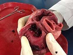
![Frontal section showing papillary muscles attached to the tricuspid valve on the right and to the mitral valve on the left via chordae tendineae.[8]](/images/5a/ab/5aabb8334721d06baf53eaa7c89d587700a9dbefdd3d33737bc25b6d9a2a4618.jpg)
Physiology and Function
The Cardiac Cycle and Blood Flow
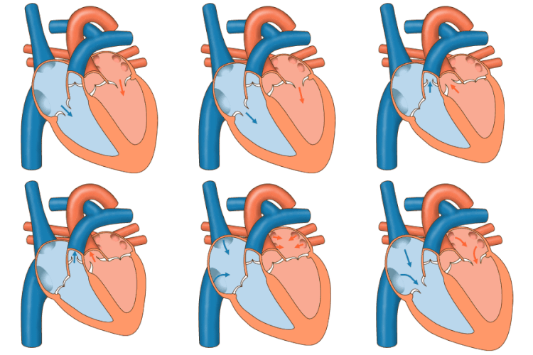
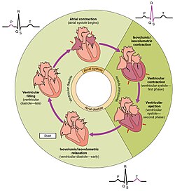
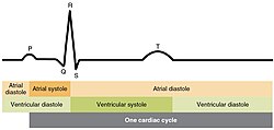
The cardiac cycle refers to the complete sequence of events in one heartbeat, involving the contraction (systole) and relaxation (diastole) of the heart muscle. This process is tightly coordinated to ensure that blood moves from the atria to the ventricles and then out to the lungs and body.
Electrical Conduction
The heart generates its own electrical impulses to trigger contractions. This conduction system ensures the atria contract first, followed by the ventricles. The process involves a prepotential—a slow influx of sodium ions that reaches a threshold to initiate spontaneous depolarization and contraction, meaning the heart cells have no resting potential.
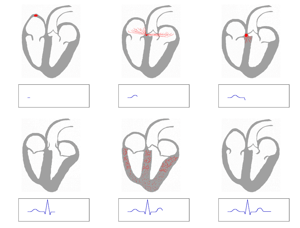
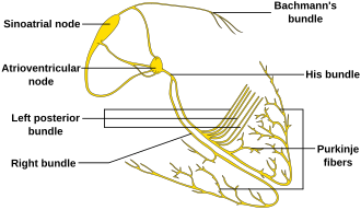
![The prepotential is due to a slow influx of sodium ions until the threshold is reached followed by a rapid depolarisation and repolarisation. The prepotential accounts for the membrane reaching threshold and initiates the spontaneous depolarisation and contraction of the cell; there is no resting potential.[8]](/images/b4/81/b481519959b18718bc50ce0a501a484875b540bb3fa120262c4bbcbb73fec6d7.jpg)
Heart Rate Dynamics
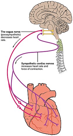
Heart rate varies significantly based on age and physical condition. While adults typically rest between 60–100 bpm, newborns may have resting rates around 129 bpm. Athletes often exhibit resting rates below 60 bpm due to increased heart efficiency. During intense exercise, heart rates can climb to 150 bpm, with maximum limits typically reaching 200 to 220 bpm.
Clinical Significance and Diagnosis
Cardiovascular Diseases
Heart diseases are largely noncommunicable and often linked to lifestyle factors and aging. Common categories include:
- Ischemic Heart Disease: Reduced blood flow to the heart muscle.
- Heart Failure: The inability of the heart to pump sufficient blood.
- Cardiomyopathies: Diseases of the heart muscle itself.
- Valvular Heart Disease: Malfunction of the heart valves.
- Cardiac Arrhythmias: Irregular heartbeats.
- Channelopathies: Genetic disorders affecting ion channels, such as Short QT syndrome.
Diagnostic Tools
Because heart disease often presents without obvious symptoms—though chest pain or shortness of breath may occur—doctors use several diagnostic methods:
- Medical History: Reviewing lifestyle and genetic risks.
- Auscultation: Using a stethoscope to listen to heart sounds caused by valve closures.
- Electrocardiogram (ECG): Recording the electrical activity of the heart.
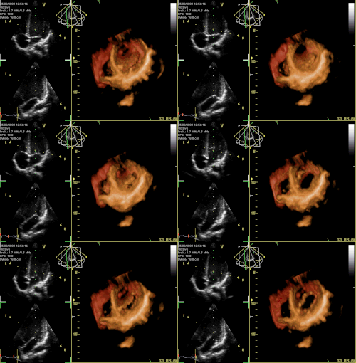
- Echocardiogram: Using ultrasound to visualize the heart's structure and movement.
Comparative Anatomy
![Development of the human heart during the first eight weeks (top) and the formation of the heart chambers (bottom). In this figure, the blue and red colors represent blood inflow and outflow (not venous and arterial blood). Initially, all venous blood flows from the tail/atria to the ventricles/head, a very different pattern from that of an adult.[8]](/images/ef/92/ef92c602fc642242157780d158899554817db430c0afed790c620cda27eb1751.jpg)
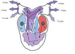
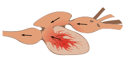
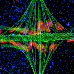
The structure of the heart varies across the animal kingdom based on the organism's respiratory needs:
| Animal Group | Heart Structure | Key Characteristic |
|---|---|---|
| Humans/Mammals | 4 Chambers | Complete separation of oxygenated and deoxygenated blood. |
| Amphibians | 3 Chambers | Single ventricle where some blood mixing occurs. |
| Fish | 2 Chambers | Simple flow: sinus venosus, atrium, ventricle, and outflow tract. |
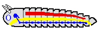
| Arthropods | Tube-like | Dorsal vessel extending across the body. |
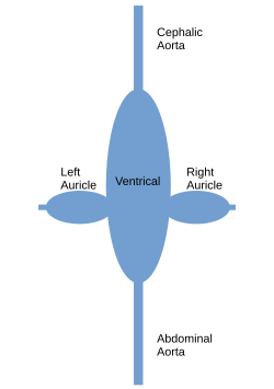
| Cephalopods | Multiple Hearts | Specialized systemic and branchial hearts. |
Frequently Asked Questions
![The x-axis reflects time with a recording of the heart sounds. The y-axis represents pressure.[8]](/images/2b/59/2b59fef6e06a3dd9eb50f6b52a11bbb3b3f05cf2bdf73e5e26aa6d339693a943.jpg)


What is the difference between levocardia and dextrocardia?
Levocardia is the normal condition where the heart is slightly offset to the left side of the chest. Dextrocardia is a rare congenital disorder where the heart is offset to the right.
Why do athletes have lower resting heart rates?
Athletes often have larger, stronger heart muscles due to regular exercise. This allows their heart to pump a greater volume of blood with each beat, meaning it needs to beat fewer times per minute to maintain the same cardiac output.
What causes the sounds a doctor hears through a stethoscope?
The characteristic "lub-dub" heart sounds are caused by the closing of the heart valves as blood is pushed through the chambers.
What are the primary risk factors for cardiovascular disease?
The most significant risk factors include smoking, being overweight, lack of physical exercise, high cholesterol, high blood pressure, and poorly managed diabetes.
How does the heart's electrical system work without a resting potential?
The heart uses a prepotential, which is a slow influx of sodium ions. Once a specific threshold is reached, it triggers rapid depolarization and repolarization, initiating the spontaneous contraction of the heart muscle.