Ventricles: Anatomy, Function, and Hemodynamics of the Heart's Lower Chambers
The heart is a sophisticated muscular pump, and its most powerful components are the ventricles. These two large chambers, located at the bottom of the heart, are responsible for collecting blood from the upper chambers (atria) and expelling it with enough force to reach the lungs and the rest of the body. While they work in tandem, the two ventricles are specialized in structure and pressure to handle very different circulatory demands.
In humans, the heart operates as a double circulatory system. The right ventricle manages the pulmonary circulation, sending blood to the lungs for oxygenation, while the left ventricle manages the systemic circulation, pumping oxygen-rich blood through the aorta to the peripheral beds of the entire body.
Key Facts
- Primary Role: Ventricles are the main pumping chambers that expel blood to the lungs and body.
- Structural Difference: The left ventricle has significantly thicker walls than the right to generate higher systemic pressure.
- Cardiac Cycle: They operate via systole (contraction/pumping) and diastole (relaxation/filling).
- Output: A healthy resting adult heart typically pumps approximately 5 liters of blood per minute.
- Pressure Gap: The left ventricle handles a workload roughly five times greater than that of the right ventricle.
Anatomical Structure and Shape
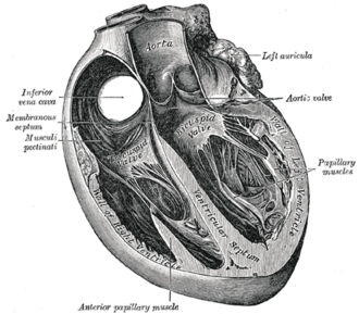
Ventricles are characterized by thicker walls than the atria, as they must generate the high pressures required to move blood over long distances. The inner walls feature irregular muscular columns known as trabeculae carneae. Among these are the papillary muscles, which anchor the chordae tendinae to the mitral and tricuspid valves, preventing them from collapsing during contraction.
The Right Ventricle
The right ventricle is triangular in shape and crescent-like in cross-section. It consists of two main components: the sinus (the inflow area from the tricuspid valve) and the conus (the outflow pouch, or conus arteriosus, leading to the pulmonary artery). It is separated from the left ventricle by the interventricular septum, a muscular wall that bulges into the right ventricular cavity.
The Left Ventricle
The left ventricle is longer, more conical, and nearly circular in transverse section. It forms the apex (the bottom tip) of the heart. Because it must overcome high aortic pressure to deliver blood to the entire body, its muscular walls are three to six times thicker than those of the right ventricle by young adulthood.
Cardiac Function and Hemodynamics
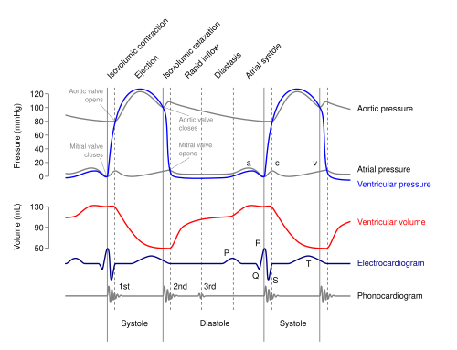
The movement of blood through the ventricles is governed by the cardiac cycle, which alternates between two primary phases:
- Systole: The phase of contraction where the ventricles pump blood out of the heart.
- Diastole: The phase of relaxation where the ventricles fill with blood from the atria.
The left ventricle receives oxygenated blood from the left atrium via the mitral valve and forces it through the aortic valve into the aorta. This process requires rapid relaxation to fill quickly and powerful contraction to overcome the pressure of the systemic arteries. Conversely, the right ventricle receives deoxygenated blood via the tricuspid valve and pumps it through the pulmonary valve into the pulmonary artery.
Pumping Volume and Performance
Cardiac performance is measured using several volumetric parameters. Stroke Volume (SV) is the amount of blood pumped per beat, while Ejection Fraction (Ef) represents the percentage of blood pumped out of the ventricle relative to the total amount it held at the end of diastole.
While a resting adult typically pumps 5 L/min, this capacity can increase significantly during exertion, reaching 25 L/min in non-athletes and up to 45 L/min in Olympic-level athletes.
Ventricular Metrics and Pressures
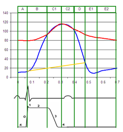
Clinical assessment of the ventricles involves measuring dimensions and pressures to ensure the heart is functioning efficiently. A significant difference between aortic and left ventricular pressure, for example, can indicate aortic stenosis.
| Measure | Right Ventricle | Left Ventricle |
|---|---|---|
| End-Diastolic Volume (EDV) | 144 mL (± 23 mL) | 142 mL (± 21 mL) |
| End-Systolic Volume (ESV) | 50 mL (± 14 mL) | 47 mL (± 10 mL) |
| Stroke Volume (SV) | 94 mL (± 15 mL) | 95 mL (± 14 mL) |
| Ejection Fraction (Ef) | 66% (± 6%) | 67% (± 4.6%) |
| Systolic Pressure (mmHg) | 15–30 | 100–140 |
| Diastolic Pressure (mmHg) | 3–8 | 3–12 |
Clinical Significance
Dysfunction in the ventricles can lead to severe medical conditions. Arrhythmias, such as ventricular fibrillation, are the most common cause of cardiac arrest and sudden death. Other structural issues include ventricular septal defects (holes in the wall between ventricles) or atrioventricular septal defects, which disrupt the normal flow of blood between the heart's chambers.
Frequently Asked Questions
Why is the left ventricle thicker than the right?
The left ventricle must pump blood to the entire body (systemic circulation), which requires much higher pressure than the right ventricle, which only needs to pump blood a short distance to the lungs (pulmonary circulation).
What is the difference between systole and diastole?
Systole is the period when the ventricular muscles contract to push blood out of the heart, while diastole is the period when the muscles relax, allowing the chambers to fill with blood again.
What does the ejection fraction measure?
The ejection fraction is a percentage that represents how much blood the ventricle pumps out with each contraction compared to the total amount of blood present in the ventricle at the end of its filling phase.
What is the interventricular septum?
The interventricular septum is the thick muscular wall that separates the right ventricle from the left ventricle, preventing the mixing of oxygenated and deoxygenated blood.
What is the role of the papillary muscles?
Papillary muscles are specialized muscles on the inner ventricular walls that attach to the heart valves via chordae tendinae, ensuring the valves do not prolapse or leak during the high-pressure contraction of systole.