Photoreceptor Cells: The Biological Engines of Vision
At the back of the human eye lies the retina, a complex layer of tissue that serves as the interface between light and the brain. The primary drivers of this system are photoreceptor cells, specialized neuroepithelial cells capable of visual phototransduction. This is the biological process of converting visible electromagnetic radiation (light) into electrical signals that the nervous system can interpret as sight.
By absorbing photons, these cells trigger changes in their membrane potential, effectively translating the physical energy of light into a biological language. While most people are familiar with rods and cones, modern science has identified a third type of photoreceptor that manages functions beyond simple image formation.
Key Facts
- Three Types: Mammalian eyes contain rods, cones, and intrinsically photosensitive retinal ganglion cells (ipRGCs).
- Rods vs. Cones: Rods handle dim-light (scotopic) vision, while cones handle bright-light (photopic) vision and color.
- Cell Count: The human retina contains approximately 120 million rods and 6 million cones.
- The Fovea: The center of the retina, known as the fovea, contains only cones and provides the highest visual acuity.
- Unique Signaling: Unlike most sensory cells, photoreceptors hyperpolarize (become more negative) when stimulated by light.
Types of Photoreceptor Cells
Rods and Cones
Rods and cones are the classic photoreceptors responsible for forming images. Rods are extremely sensitive to light, capable of registering a single photon, making them essential for vision in low-light conditions. However, they provide low spatial resolution and achromatic (black and white) vision. Cones require more light to activate but provide high visual acuity and the ability to perceive color.
Humans possess three classes of cones—S, M, and L—each with a different spectral sensitivity (the range of wavelengths they prefer). For instance, S-cones peak at approximately 420 nm. This diversity allows the visual system to transduce a full spectrum of colors.
![Normalized human photoreceptor absorbances for different wavelengths of light[3]](/images/cd/a6/cda6b3b4dc53d85c11eb1e27d89d9cbeaadf9dc7158e2a7f5c7885ede97491a7.webp)
Intrinsically Photosensitive Retinal Ganglion Cells (ipRGCs)
Discovered in the 1990s, ipRGCs are a small subset (1–3%) of retinal ganglion cells. Unlike rods and cones, they contain a light-sensitive protein called melanopsin. These cells do not contribute to sight directly; instead, they regulate the pupillary light reflex and the entrainment of the circadian rhythm (the body's internal 24-hour clock).
Anatomy and Histology
Rods and cones share a similar basic structure, organized into specific segments that facilitate the movement of signals toward the brain.
- Outer Segment: Modified cilia containing disks filled with opsin (the light-absorbing molecule) and voltage-gated sodium channels. This is where light absorption occurs.
- Inner Segment: Packed with mitochondria to provide ATP (energy) for the sodium-potassium pump.
- Cell Body: Contains the cell's organelles.
- Axon Terminal: The point where the neurotransmitter glutamate is released to bipolar cells.
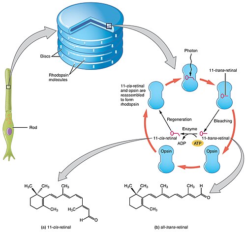
The specific pigment depends on the cell type: in rods, the combination of opsin and retinal is called rhodopsin; in cones, these are called photopsins.
The Retinal Mosaic and Distribution
The arrangement of these cells across the retina is known as the retinal mosaic. This distribution is not uniform. The fovea centralis, located directly behind the lens, is densely packed with cones and contains no S-cones, resulting in the highest resolution of vision. Conversely, the blind spot is an area where no photoreceptors exist because the ganglion cell fibers gather to form the optic nerve.
![Distribution of rods and cones along a line passing through the fovea and the blind spot of a human eye[7]](/images/b2/d3/b2d3591b43dc9e85aff3ff9548175bb30c48e4e4663f38d8312fe694e4a08afb.webp)
The Phototransduction Cascade
The process of converting light into a neural signal is a complex chain reaction called the phototransduction cascade. Interestingly, photoreceptors are depolarized in the dark (the "dark current"), meaning they continuously release glutamate when no light is present.
- Activation: A photon hits the opsin, causing an isomeric change in the retinal molecule.
- First Amplification: The activated opsin triggers a G protein called transducin. One opsin can activate about 100 transducins.
- Second Amplification: Transducin activates phosphodiesterase (PDE), which hydrolyzes cGMP into 5' GMP. One PDE can hydrolyze about 1,000 cGMP molecules.
- Channel Closure: The drop in cGMP levels causes sodium (Na) ion channels to close.
- Hyperpolarization: The cell becomes hyperpolarized, reducing the release of glutamate to the bipolar cells.
This unique system reduces "noise" and allows for massive amplification, ensuring that even a single photon can trigger a signal to the brain.
Comparison of Rods and Cones
| Feature | Rods | Cones |
|---|---|---|
| Vision Type | Scotopic (Low light) | Photopic (Bright light) |
| Sensitivity | Very high (single photon) | Lower (requires direct light) |
| Visual Acuity | Low | High |
| Color Vision | Achromatic | Trichromatic (S, M, L) |
| Fovea Presence | Absent | Concentrated |
| Response Speed | Slow | Fast |
| Quantity | ~120 Million | ~6 Million |
Frequently Asked Questions
What happens if a person loses their rod cells?
The loss of rod cells typically results in night blindness, as the eye can no longer function effectively in scotopic (low-light) conditions.
What happens if a person loses their cone cells?
The loss of cone cells can lead to legal blindness and a total loss of color vision, as cones are responsible for high-resolution sight and color perception.
Why do photoreceptors hyperpolarize instead of depolarize?
This mechanism reduces sensory noise. Because the cell is already depolarized in the dark, the random opening or closing of a few channels doesn't trigger a signal; only the coordinated closing of many channels via light absorption creates a meaningful signal.
How do ipRGCs help people who are otherwise blind?
Because ipRGCs are located in the inner retina and are separate from rods and cones, some individuals who have lost their outer retina still maintain circadian rhythms and pupillary reflexes, as their ipRGCs remain functional.
What is the role of glutamate in the retina?
Glutamate is the neurotransmitter released by photoreceptors to signal bipolar cells. In the dark, it is released continuously; when light is absorbed, the release of glutamate decreases, which the bipolar cells interpret as a visual stimulus.