Retina Anatomy, Function, and Clinical Significance
The retina is the light-sensitive inner lining of the eye, acting as the critical interface between the physical world and the brain's visual processing centers. In adult humans, the retina covers approximately 72% of a sphere with a 22 mm diameter. It transforms incoming light into electrical signals, performing significant pre-processing before the information ever reaches the optic nerve.
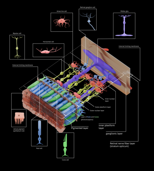
The complexity of the retina lies in its layered architecture, which allows it to filter raw visual data into recognizable shapes, edges, and movement. This biological sensor is composed of millions of specialized cells, including photoreceptors and various nerve layers that refine the visual image.
Key Facts
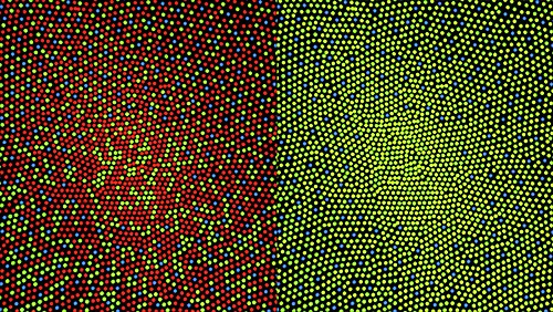
- Photoreceptor Count: The human retina contains roughly 7 million cones and between 75 to 150 million rods.
- The Blind Spot: The optic disc is a 3 mm oval area lacking photoreceptors where the optic nerve exits the eye.
- Central Vision: The fovea is a specialized pit responsible for sharp central vision, though it is less sensitive to light due to a lack of rods.
- Data Compression: While there are over 130 million receptors, only about 1.2 million axons travel through the optic nerve, indicating massive internal data processing.
- Foveal Capacity: The fovea occupies only 0.01% of the visual field but utilizes 10% of the optic nerve's axons.
Retinal Structure and Layers
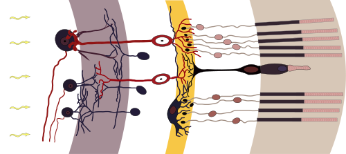
The vertebrate retina is organized into 10 distinct layers. The arrangement is considered "inverted," meaning light must pass through several transparent nerve layers before reaching the photoreceptors at the back.
The Visual Pathway
Visual signals follow a specific flow: they begin as raw outputs from photoreceptors (rods and cones), move to bipolar cells, then to ganglion cells, and finally travel through the optic chiasm to the lateral geniculate nucleus (LGN) and the V1 cortex of the brain.
Specialized Regions
- Macula: Located temporal to the optic disc, this area is central to high-resolution vision.
- Fovea: The center of the macula, optimized for detail. Humans and primates have one fovea, whereas some birds are bifoviate and some mammals possess a visual streak instead.
- Ora Serrata: The farthest edge of the retina.
- Peripheral Retina: The area extending from the central retina (roughly 6 mm around the fovea) to the ora serrata.
![Distribution of rods and cones along a line passing through the fovea and the blind spot of a human eye[13]](/images/0e/c8/0ec86c670d7b703d38a0e080b1499acec92366e6b199127724d5d8e019915be9.webp)
Optical Coherence Tomography (OCT) Analysis
Optical Coherence Tomography (OCT) is a non-invasive imaging technique used to visualize the retinal layers with high resolution. OCT can distinguish between hyper-reflective (bright) and hypo-reflective (dark) zones to diagnose pathology.
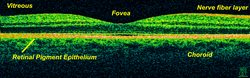
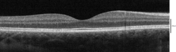
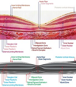
The following table summarizes the key layers identifiable via OCT and their anatomical correlates.
| OCT Layer | Anatomical Correlate | Reflectivity |
|---|---|---|
| Internal Limiting Membrane (ILM) | Müller cell endfeet | Hyper-reflective |
| Nerve Fiber Layer (NFL) | Ganglion cell axons | Variable |
| Ganglion Cell Layer (GCL) | Ganglion cell bodies | Hypo-reflective |
| Inner Plexiform Layer (IPL) | Bipolar, amacrine, and ganglion synapses | Hyper-reflective |
| Inner Nuclear Layer (INL) | Horizontal, bipolar, and amacrine cell bodies | Hypo-reflective |
| Outer Plexiform Layer (OPL) | Photoreceptor, bipolar, and horizontal synapses | Hyper-reflective |
| Outer Nuclear Layer (ONL) | Photoreceptor cell bodies | Hypo-reflective |
| Ellipsoid Zone (EZ) | Photoreceptor inner segment (mitochondria) | Very Hyper-reflective |
| RPE / Bruch's Complex | Retinal Pigment Epithelium | Variable/Hyper-reflective |
Clinical Significance and Treatment
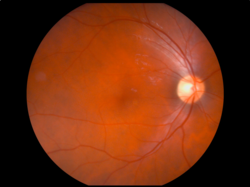
The retina's blood supply is primarily managed by the central retinal artery. Because the retina is a window into the vascular system, fundus photography can reveal the state of blood vessels, where veins typically appear darker and wider than arteries.
Management of Retinal Disease
Retinal disorders can be inherited or acquired. Modern medical management includes several modalities:
- Intravitreal Medication: Injection of corticosteroids or anti-VEGF agents directly into the eye.
- Vitreoretinal Surgery: Surgical intervention to repair physical retinal damage.
- Nutritional Support: Use of specific supplements to maintain retinal health.
- Advanced Therapies: Retinal gene therapy is an emerging, though less common, treatment modality.
Frequently Asked Questions
What is the difference between rods and cones?
Rods are highly sensitive to light and are primarily responsible for vision in low-light conditions and peripheral vision. Cones are concentrated in the fovea and are responsible for high-resolution central vision and color perception.
Why is the optic disc called the blind spot?
The optic disc is the point where the optic nerve fibers exit the eye to travel to the brain. Because there are no photoreceptors (rods or cones) located in this specific area, no light can be detected, creating a gap in the visual field.
What is the function of the fovea?
The fovea is a small pit in the center of the macula that provides the highest visual acuity. It is densely packed with cones and is responsible for tasks requiring sharp detail, such as reading.
How does the retina process information before it reaches the brain?
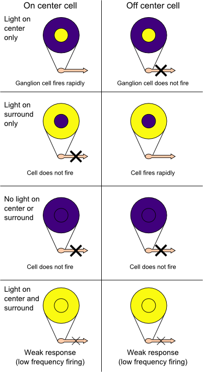
The retina performs spatial encoding through a network of bipolar, horizontal, and amacrine cells. This pre-processing identifies simple shapes, edges, and movement, significantly compressing the data from 130 million receptors down to 1.2 million optic nerve fibers.