Nictitating Membrane: The Biology and Function of the Third Eyelid
In the diverse world of animal anatomy, some species possess a remarkable ocular feature known as the nictitating membrane. Derived from the Latin word nictare, meaning "to blink," this structure acts as a transparent or translucent third eyelid. Unlike the primary upper and lower eyelids that move vertically, the nictitating membrane slides horizontally across the eyeball from the medial canthus (the inner corner of the eye) to protect and moisten the ocular surface without completely blocking vision.
Scientifically referred to as the membrana nictitans, palpebra tertia, or plica semilunaris, this membrane is often informally called a "haw." It serves as a critical adaptation for survival, allowing animals to navigate harsh environments—from the depths of the ocean to the high-speed dives of a falcon—while keeping their eyes safe and hydrated.
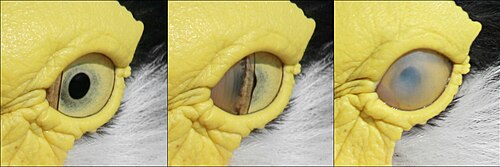
Key Facts
- Function: Protects the eye from debris and moisture loss while maintaining visibility.
- Movement: Moves horizontally across the eye, unlike standard eyelids.
- Distribution: Found in most Anura (tailless amphibians), some reptiles, birds, sharks, and specific mammals.
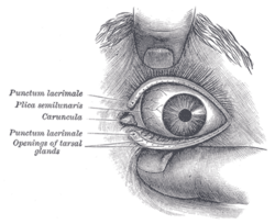
- Human Equivalent: The plica semilunaris (semilunar fold) is the vestigial remnant of this membrane in humans.
- Tear Production: In some animals, the attached Harder's gland (nictitans gland) can produce up to 50% of the tear film.
Distribution Across Species
The prevalence of the nictitating membrane varies significantly across the animal kingdom. It is common in fish, amphibians, reptiles, and birds. Among mammals, fully developed membranes are found in species such as cats, beavers, polar bears, seals, sheep, and aardvarks.
In primates, the membrane is rare. While lemurs and lorisoid primates possess fully developed versions, most other primates do not. In humans, the structure has evolved into a vestigial remnant known as the plica semilunaris, located in the corner of the eye.
Comparative Anatomy Summary
| Animal Group | Status of Membrane | Examples |
|---|---|---|
| Amphibians | Fully Developed | Most Anura (excluding Hymenochirus, Pseudhymenochirus, and Pipa) |
| Birds | Fully Developed | Peregrine falcons, Woodpeckers |
| Marine Animals | Fully Developed | Sharks, Seals, Manatees |
| Mammals | Variable | Cats, Polar bears, Aardvarks |
| Primates | Mostly Vestigial | Humans (plica semilunaris), Lemurs (full) |
Specialized Functions and Adaptations
The nictitating membrane is not a one-size-fits-all tool; it has evolved to meet the specific environmental challenges of different species.
Environmental Protection
For diving animals like sea lions, the membrane is activated on land to clear sand and debris. In beavers and manatees, the transparent membrane protects the eye while submerged. Crocodiles use it for underwater protection, though it can hinder their ability to focus while submerged. Polar bears utilize the membrane to prevent snow blindness by shielding the eye from ultraviolet radiation.
High-Impact and High-Speed Utility
Birds exhibit sophisticated control over their third eyelid. Peregrine falcons, which dive at speeds of 200 mph (320 km/h), blink their nictitating membranes repeatedly to spread moisture and clear debris. Woodpeckers employ a unique reflex, tightening the membrane a millisecond before their beak hits a tree trunk to prevent retinal injury caused by the impact vibration.
Predation and Defense
Sharks use the membrane to protect their eyes during attacks on prey. Similarly, birds of prey use the membrane to shield their eyes from their chicks during feeding.
Clinical Significance and Vestigiality
In domestic animals like cats and dogs, the nictitating membrane lacks significant muscle fibers and is typically not visible. If the membrane becomes chronically visible, it is often an indicator of poor health or illness. However, it can be observed in healthy animals during sleep or by applying gentle pressure to the eyeball, which triggers a reflex response.
In certain dog breeds, the gland associated with the third eyelid can prolapse, leading to a medical condition known as cherry eye.
Frequently Asked Questions
Do humans have a third eyelid?
Humans do not have a functional third eyelid, but we possess a vestigial remnant called the plica semilunaris, which is the small fold of conjunctiva in the inner corner of the eye.
How does the nictitating membrane differ from normal eyelids?
While standard eyelids move vertically to close the eye, the nictitating membrane moves horizontally across the eyeball.
What is "cherry eye" in dogs?
Cherry eye is a condition where the gland of the third eyelid prolapses, becoming visible as a red mass in the corner of the dog's eye.
Why do woodpeckers use their nictitating membrane?
Woodpeckers close the membrane a millisecond before impact with a tree to protect their retinas from the shock of the strike.
Can the nictitating membrane be used in scientific research?
Yes, the reflex response of the nictitating membrane to stimuli (such as a puff of air) is frequently used in classical conditioning experiments, particularly in rabbits.