DNA: The Blueprint of Life
Every living organism, from the smallest bacterium to the largest whale, relies on a complex molecular instruction manual to grow, survive, and reproduce. This manual is DNA (deoxyribonucleic acid), a molecule that carries the genetic instructions necessary for the development and functioning of all known living organisms. By storing information in a precise chemical code, DNA ensures that biological traits are passed from one generation to the next through the process of heredity.
The Structure of DNA: The Double Helix
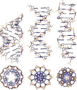
At its most fundamental level, DNA is composed of two long strands that wind around each other, creating a shape known as a double helix. The most common form of this structure is called B-DNA. These two strands are held together by hydrogen bonds between nitrogenous bases, which act like the rungs of a twisting ladder.
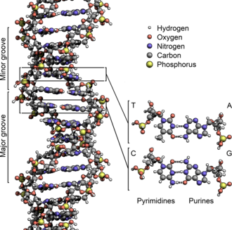
To understand the complexity of DNA, it helps to look at it through a simplified lens before diving into the chemistry.
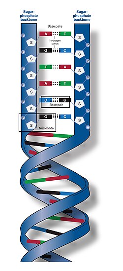
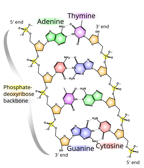
The chemical backbone of DNA consists of alternating sugar and phosphate groups. Each strand has a specific directionality, defined by its ends: one end has an exposed 5' phosphate group, and the other has an exposed 3' hydroxyl group (—OH). The "rungs" of the ladder are made of four nucleobases: adenine (A), thymine (T), cytosine (C), and guanine (G). These bases follow strict pairing rules: adenine always pairs with thymine, and cytosine always pairs with guanine.
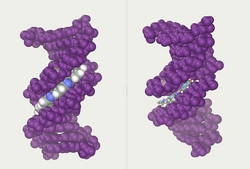
In a section of DNA, these bases lie horizontally between the two spiraling strands, protecting the genetic code within the center of the helix.
![A section of DNA. The bases lie horizontally between the two spiraling strands[15] (animated version).](/images/24/2a/242a6adcd2c36e0e7d881846278cf04a3ef81217f445f8943d27b96f1f40dfaa.gif)
The twisting of the strands creates gaps of different sizes known as major and minor grooves. These grooves are not just structural; they serve as critical binding sites for proteins and dyes, such as the Hoechst stain dye 33258, which allows scientists to visualize DNA.
From Nucleotides to Genomes: The Hierarchy of Genetic Information
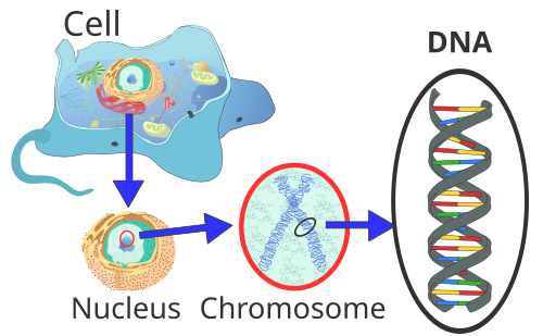
DNA is organized in a hierarchical system that allows a massive amount of information to fit inside a microscopic cell. This organization moves from the smallest chemical unit to the entire genetic library of an organism.
- Nucleotides: The basic building blocks of DNA, consisting of a sugar, a phosphate, and a base.
- Genes: Specific sequences of DNA base pairs that encode instructions for a particular function, such as producing a protein.
- Chromosomes: Long, packaged strands of DNA. A human cell contains 22 pairs of homologous chromosomes (chromosomes that are similar in shape and gene content) and one pair of sex chromosomes (XX for females, XY for males).
- Genome: The complete set of genetic material in an organism, including both nuclear DNA and mitochondrial DNA (DNA found in the cell's energy-producing organelles).
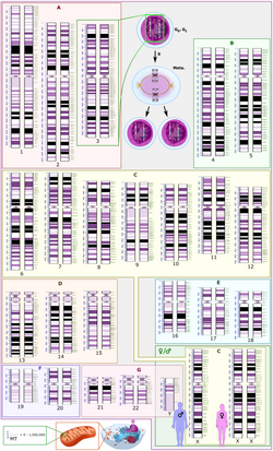
In eukaryotes (organisms with a distinct nucleus), the majority of this DNA is housed within the nucleus, tightly coiled into chromosomes to prevent tangling and damage.
How DNA Functions: Replication and Packaging
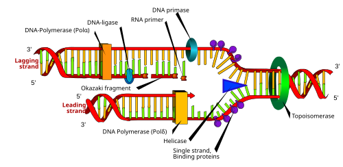
For a cell to divide, it must first copy its entire genetic library. This process is called DNA replication. It begins when enzymes called helicase and topoisomerase unwind the double helix. Then, DNA polymerase creates new complementary strands. While the "leading strand" is built continuously, the "lagging strand" is created in short, discontinuous segments called Okazaki fragments, which are eventually joined together by DNA ligase.
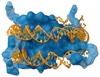
Because DNA strands are incredibly long, they cannot simply float loosely in the nucleus. They wrap around proteins called histones. The basic amino acids in histones bind to the acidic phosphate groups of the DNA, packaging the molecule into a compact structure.
Alternative Structures and Chemical Modifications
While the B-DNA double helix is the most common form, DNA is highly flexible and can adopt other shapes depending on the environment and sequence. These include A-DNA and Z-DNA (a left-handed helix).
In some cases, DNA can form a quadruplex—a four-stranded structure often found in telomere repeats (the protective caps at the ends of chromosomes). These structures are often stabilized by potassium ions.
![DNA quadruplex formed by telomere repeats. The looped conformation of the DNA backbone is very different from the typical DNA helix. The green spheres in the center represent potassium ions.[62]](/images/02/77/0277c3de7b21842e8a34d2805f12a443d7b02f1e2a7c77142d915db8274b4a36.png)
DNA can also undergo chemical modifications. Methylation, such as the conversion of cytosine to 5-methylcytosine, is a key part of epigenetics—the study of how behaviors and environment cause changes that affect the way genes work without changing the DNA sequence itself.
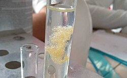
When DNA is extracted in an impure state, such as from an orange, it often appears as white, stringy clumps.
However, DNA can also be damaged by external factors. For example, benzo[a]pyrene, a mutagen found in tobacco smoke, can form a covalent adduct (a chemical bond) with DNA, potentially leading to mutations.
![A covalent adduct between a metabolically activated form of benzo[a]pyrene, the major mutagen in tobacco smoke, and DNA[82]](/images/58/c3/58c3ff4435feca420843a52ee079cd0d16ad29064142a4b88edaed73e80aad3f.png)
Interactions with Proteins and Genetic Recombination
DNA does not act alone; it interacts with various proteins to regulate gene expression. Transcription factors, such as the lambda repressor, bind to specific DNA targets to turn genes on or off.
![The lambda repressor helix-turn-helix transcription factor bound to its DNA target[117]](/images/8d/2d/8d2d68f165564e7dbf2ca962250e3c6e4f1c1c1c695c82903736152489c00f1e.png)
Other enzymes, like restriction enzymes (e.g., EcoRV), act as molecular scissors that cut DNA at specific sequences, a tool widely used in genetic engineering.
![The restriction enzyme EcoRV (green) in a complex with its substrate DNA[121]](/images/8f/fb/8ffbf9773b794489f5586e9dad9fcd8fb1f4c5297f7cfeb34da00c69fd7577a5.png)
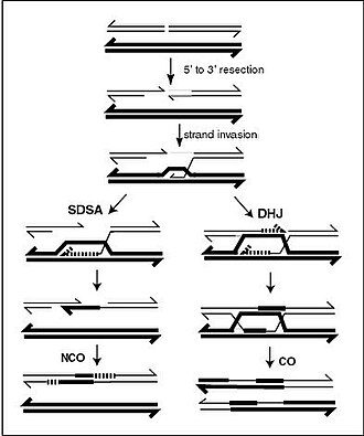
During meiosis (the cell division that produces sperm and egg cells), genetic recombination occurs. This process involves double-strand breaks and strand invasion between homologous chromosomes. This can result in a "crossover" (CO) via the Double Holliday Junction (DHJ) model or a "non-crossover" (NCO) via the Synthesis Dependent Strand Annealing (SDSA) model, ensuring genetic diversity in offspring.
The Future of DNA: Technology and Nanotechnology
Beyond biology, scientists are using the molecular recognition properties of DNA to build nanoscale structures. This field, known as DNA nanotechnology, allows for the self-assembly of precise shapes and machines at the atomic level.
![The DNA structure at left (schematic shown) will self-assemble into the structure visualized by atomic force microscopy at right. DNA nanotechnology is the field that seeks to design nanoscale structures using the molecular recognition properties of DNA molecules.[171]](/images/30/ed/30ed5ded23b9f64792fb26746e361120c11b36a9b5633bd01bb867a888a74aae.png)
Other modern applications include:
- DNA Profiling: Using unique genetic markers for forensic identification.
- Bioinformatics: Using computer science to analyze vast genomic datasets.
- Information Storage: Exploring the use of DNA as a high-density digital storage medium.
The History of Discovery
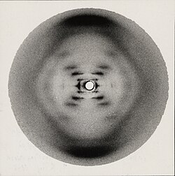
The discovery of the double helix in 1953 was a landmark moment in science. While Francis Crick and James Watson are often credited with the model, their work relied heavily on the X-ray diffraction data produced by Rosalind Franklin and Raymond Gosling.
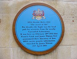
The discovery is commemorated in Cambridge, England, with a plaque outside The Eagle pub, where Crick and Watson famously announced their findings.
The most critical piece of evidence was "Photo 51," an X-ray diffraction pattern that revealed the helical nature of the molecule.
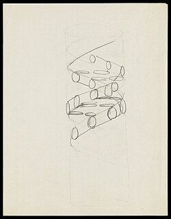
Early sketches, such as those by Francis Crick, helped translate these mathematical patterns into the physical model we recognize today.
Summary of DNA Key Concepts
The following table summarizes the essential components and processes of DNA.
| Term | Definition/Function | Key Example/Component |
|---|---|---|
| Nucleotide | The basic building block of DNA | Sugar, Phosphate, Base |
| Base Pair | Complementary nitrogenous bases | A-T and C-G |
| Gene | A segment of DNA encoding a function | Protein-coding sequences |
| Chromosome | Packaged DNA strand | Human karyotype (23 pairs) |
| Replication | The process of copying DNA | DNA Polymerase, Helicase |
| Histone | Protein used for DNA packaging | Nucleosomes |