Digestive System: Anatomy and Function
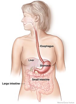
The digestive system, also known as the alimentary system, is a complex network of organs designed to break down food into nutrients, which the body uses for energy, growth, and cell repair. This process involves a coordinated sequence of mechanical and chemical actions, starting from the moment food enters the mouth and ending with the elimination of waste.
From the secretion of enzymes in the salivary glands to the absorption of critical vitamins in the small intestine, every component plays a specialized role in maintaining homeostasis.
Key Facts
- Saliva Production: The submandibular glands produce approximately 70% of the oral cavity's saliva.
- Starch Digestion: Up to 30% of initial starch digestion occurs in the mouth via amylase.
- Esophagus Length: The average length of the esophagus is 25 cm, though this varies by height.
- Small Intestine Transit: Food residues typically take an average of 4 hours to transit the small intestine, with complete emptying occurring after about 8.6 hours.
- Blood Flow: Blood flow to the digestive tract peaks 20–40 minutes after a meal and lasts for 1.5 to 2 hours.
The Upper Gastrointestinal Tract
The Mouth and Salivary Glands
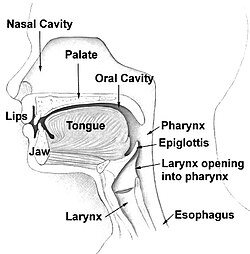
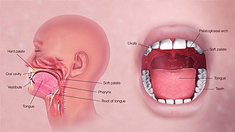
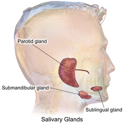
Digestion begins in the mouth, where food is mechanically broken down by teeth and moistened by saliva. Saliva transforms food into a smooth bolus, facilitating its passage into the esophagus. This process is supported by three pairs of main exocrine salivary glands:
- Parotid glands: The largest glands, producing primarily serous secretion.
- Submandibular glands: Located under the jaw, these produce both mucus and serous fluid, including lingual lipase.
- Sublingual glands: Located under the tongue, producing mainly mucous secretions.
Chemical Digestion in the Mouth
Saliva contains essential enzymes that initiate chemical breakdown. Amylase targets carbohydrates, breaking starch into simple sugars like maltose and dextrose. Lipase begins the breakdown of fats; this is particularly critical for infants whose pancreatic lipase has not yet fully developed.
The Esophagus
The esophagus (or foodpipe) is a muscular tube approximately 25 cm long that transports the bolus from the pharynx to the stomach. It passes through the posterior mediastinum of the thorax and enters the stomach via the esophageal hiatus—an opening in the thoracic diaphragm located at the level of the tenth thoracic vertebra (T10).
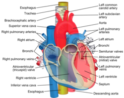
The Stomach and Mid-Digestive Organs
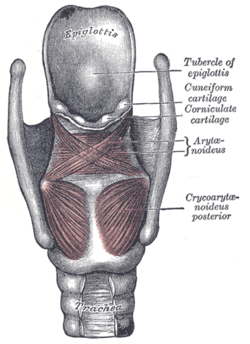
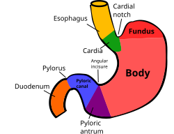
Stomach Function and Vitamin B12
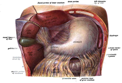
The stomach is not only a site for mechanical churning but also a critical center for nutrient preparation. Parietal cells in the fundus of the stomach produce intrinsic factor, a glycoprotein essential for the absorption of vitamin B12 (cobalamin). To protect the acid-sensitive vitamin B12 from gastric acid, it first binds to transcobalamin I (haptocorrin) secreted by salivary glands. Once it reaches the neutral environment of the duodenum, pancreatic enzymes release the vitamin, allowing it to bind with intrinsic factor for absorption in the ileum.
Supporting Organs: Liver, Gallbladder, and Pancreas
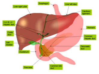
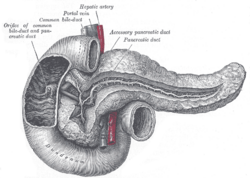
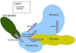
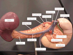
The liver and gallbladder manage bile, which is necessary for fat digestion. The pancreas secretes further lipase and other enzymes into the duodenum to continue the chemical breakdown of nutrients.
The Lower Gastrointestinal Tract
The Small Intestine
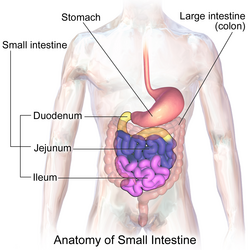
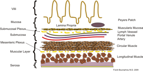
The small intestine is the primary site for nutrient absorption. The transit time for food residues is an average of 4 hours, though complete emptying takes roughly 8.6 hours. Blood supply to this region is provided by the superior mesenteric artery, which serves the midgut, including the distal two-thirds of the duodenum, jejunum, ileum, cecum, appendix, ascending colon, and the proximal two-thirds of the transverse colon.
The Large Intestine
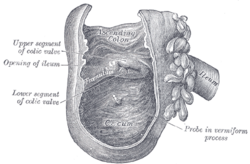
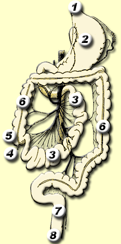
The lower tract begins at the cecum, leading into the large intestine (colon), where water is absorbed and waste is prepared for excretion.
Clinical Significance and History
Digestive System Health
Cancers of the digestive system remain a significant health challenge. In 2023, the United States saw an estimated 348,840 new cases and 172,010 deaths. The highest mortality rates were associated with cancers of the colon and rectum, followed by the pancreas, liver, esophagus, and stomach. Research indicates that abnormally high levels of bile acids in specific regions may act as carcinogens.
Historical Milestones
| Era/Year | Contributor | Discovery/Contribution |
|---|---|---|
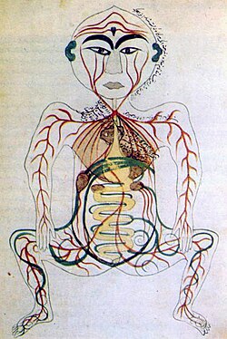
| 16th Century | Leonardo da Vinci & Andreas Vesalius | Early anatomical drawings of the stomach and intestines. |
| 1823 | William Prout | Discovered hydrochloric acid in gastric juice. |
| 1825 | William Beaumont | Observed the churning motion of the stomach in a living patient. |
| 1895 | Ivan Pavlov | Linked gastric secretion to neurologic reflexes via the vagus nerve. |
| 1902 | Ernest Starling | Discovered the hormone secretin. |
| 1983 | Marshall & Warren | Identified Helicobacter pylori as a cause of ulcers. |

Frequently Asked Questions
How much of starch digestion happens in the mouth?
Saliva contains the enzyme amylase, which can account for approximately 30% of the initial digestion of starch into simple sugars like maltose and dextrose.
What is the role of intrinsic factor in the stomach?
Intrinsic factor is a glycoprotein produced by parietal cells in the stomach's fundus. It is essential for the absorption of vitamin B12 in the ileum.
How long does food typically stay in the small intestine?
The average transit time is 4 hours, but it takes an average of 8.6 hours for the small intestine to empty completely.
Which artery supplies the majority of the midgut?
The superior mesenteric artery supplies the regions derived from the midgut, including the jejunum, ileum, cecum, and parts of the duodenum and colon.
What is the significance of Helicobacter pylori?
Discovered in 1983 by Barry Marshall and Robin Warren, Helicobacter pylori is a bacterium identified as a primary cause in the formation of gastric ulcers.