Ulna Anatomy: Structure, Function, and Clinical Significance
The ulna, or ulnar bone, is one of the two long bones located in the human forearm. Stretching from the elbow to the wrist, it runs parallel to the radius and is positioned on the medial side of the forearm—the same side as the little finger when the body is in the standard anatomical position. While it is longer and thinner than the radius, it serves as a critical structural component for the stability of the elbow and the movement of the forearm.
In the human body, the ulna is the smaller of the two lower arm bones, functioning similarly to how the fibula operates in the lower leg.
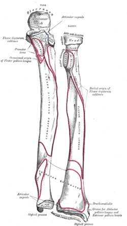
Key Facts
- Location: Medial side of the forearm (pinky-finger side).
- Primary Joints: Forms a hinge joint at the elbow and contributes to the wrist complex.
- Key Landmarks: Includes the olecranon (the point of the elbow) and the ulnar styloid process.
- Development: Ossifies from three separate centers starting around the eighth week of fetal life.
- Comparative Anatomy: In birds and dinosaurs, the ulna features "quill knobs" for secondary feather attachment.
Detailed Anatomical Structure
The ulna is broader near the elbow and gradually narrows as it approaches the wrist. Its structure is divided into three primary regions: the proximal end (near the elbow), the body (shaft), and the distal end (near the wrist).
The Proximal End (The Elbow)
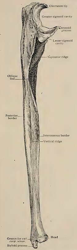
The upper portion of the ulna is characterized by two curved processes and two articular cavities that facilitate elbow movement:
- Olecranon Process: A thick, hook-like eminence that forms the point of the elbow. It fits into the olecranon fossa of the humerus, preventing hyperextension of the arm.
- Coronoid Process: A triangular projection that fits into the coronoid fossa of the humerus during forearm flexion.
- Semilunar Notch: A large, concave depression formed by the olecranon and coronoid processes, which articulates with the trochlea of the humerus to create a hinge joint.
- Radial Notch: A narrow depression on the lateral side of the coronoid process that receives the head of the radius, allowing for a pivot joint.
The Body (The Shaft)
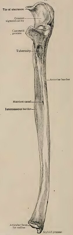
The body of the ulna is prismatic and tapers downward. It is defined by three borders and three surfaces:
- Borders: The volar (anterior) border, the dorsal (posterior) border, and the interosseous crest. The interosseous crest is particularly important as it attaches to the interosseous membrane, a fibrous sheet connecting the ulna to the radius.
- Surfaces: The volar surface is concave and serves as an origin for muscles like the flexor digitorum profundus. The dorsal surface features a longitudinal ridge (perpendicular line) and provides attachment points for the supinator and extensor muscles.
The Distal End (The Wrist)
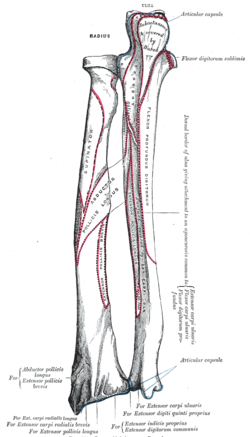
Near the wrist, the ulna terminates in two distinct eminences:
- Head of the Ulna: A rounded articular eminence that fits into the ulnar notch of the radius.
- Ulnar Styloid Process: A non-articular projection on the medial side that provides an attachment point for the ulnar collateral ligament of the wrist.
Microanatomy and Development
The ulna is a classic long bone featuring a medullary cavity enclosed by a thick wall of cortical tissue. This compact bone is thickest along the dorsal surface and the interosseous border. At the ends, the bone transitions into a network of spongy bone (trabeculae) to better distribute mechanical loads.
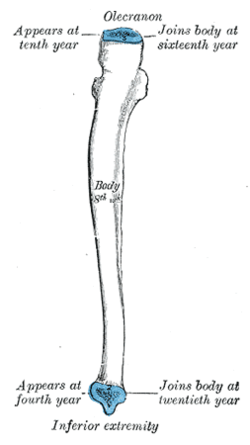
Developmentally, the ulna ossifies from three centers: the body, the wrist end, and the elbow end. Ossification begins around the eighth week of fetal development. The epiphyses (the ends of the bone) typically fuse with the body between the ages of 16 and 20.
Joints and Muscle Attachments
The ulna is central to several key articulations and serves as an anchor for numerous muscles that control the hand and arm.
| Category | Connection/Muscle | Function/Role |
|---|---|---|
| Joints | Humerus (Trochlea) | Hinge joint for elbow flexion/extension |
| Joints | Radius (Head) | Pivot joint for pronation/supination |
| Muscle | Triceps Brachii | Insertion at Olecranon; extends the forearm |
| Muscle | Brachialis | Insertion at Coronoid process; flexes the forearm |
| Muscle | Pronator Quadratus | Origin at distal shaft; rotates the forearm |
Clinical Significance
Fractures of the ulna are common and vary in severity. Two specific clinical patterns include:
- Monteggia Fracture: A fracture of the proximal third of the ulna accompanied by the dislocation of the radial head.
- Hume Fracture: A fracture of the olecranon combined with an anterior dislocation of the radial head.
Conservative management, such as casting above the elbow, is typically reserved for fractures in the distal two-thirds of the shaft that exhibit minimal displacement (less than 50%) and low angulation (less than 10°).
Comparative Anatomy in Other Animals
In four-legged mammals, the radius is usually the primary load-bearing bone, while the ulna primarily serves as a site for muscle attachment. In some species, such as horses, the ulna is partially or wholly fused with the radius.
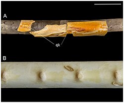
In birds and dinosaurs, the ulna is specialized for flight or balance. It provides the attachment surface for secondary feathers, leaving behind quill knobs—small bony bumps that allow paleontologists to identify feathers in fossilized remains.
Frequently Asked Questions
What is the difference between the ulna and the radius?
The ulna is the medial bone of the forearm (on the pinky side) and is primarily involved in the hinge joint of the elbow. The radius is the lateral bone (on the thumb side) and is the primary bone that rotates around the ulna to allow the palm to turn up or down.
What is the olecranon process?
The olecranon process is the large, hook-like bony projection at the top of the ulna. It forms the prominent point of the elbow and serves as the insertion point for the triceps brachii muscle.
What happens during a Monteggia fracture?
A Monteggia fracture occurs when the upper third of the ulna breaks and the head of the radius is simultaneously dislocated from its joint at the elbow.
How does the ulna function in birds?
In birds, the ulna supports the secondary flight feathers. These feathers attach to the bone at specific points called quill knobs, which are often preserved in the fossil record of dinosaurs and early birds.
At what age does the ulna finish growing?
While ossification begins in the womb, the epiphyses (growth plates) at the ends of the bone typically fuse with the main shaft between the ages of 16 and 20.