Humerus Anatomy: Structure, Function, and Clinical Significance
The humerus is the definitive long bone of the upper arm, extending from the shoulder to the elbow. It serves as the critical structural link between the scapula (shoulder blade) and the two bones of the forearm: the radius and the ulna. Its complex architecture allows for a wide range of motion at the shoulder and provides stable articulation at the elbow.
Anatomically, the humerus is divided into three primary sections: the proximal (upper) extremity, the cylindrical shaft (body), and the distal (lower) extremity. Each section features specific landmarks that serve as attachment points for muscles and conduits for vital nerves and blood vessels.
Key Facts
- Location: The single bone of the upper arm, connecting the shoulder to the elbow.
- Critical Zones: The surgical neck is a frequent site of fractures.
- Nerve Association: The radial nerve travels closely along the shaft in the spiral groove.
- Joints: Articulates with the scapula (shoulder), radius, and ulna (elbow).
- Development: One of the first bones to ossify in the embryo, used as a biometric for gestational age.
The Upper Extremity (Proximal End)
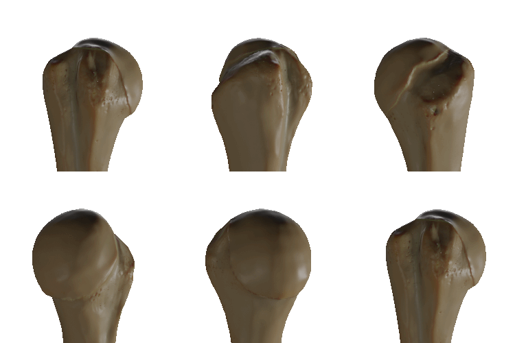
The proximal end of the humerus is designed for the high mobility of the shoulder joint. It consists of a large, rounded head that fits into the glenoid fossa of the scapula.
The Anatomical and Surgical Necks
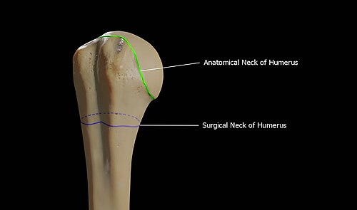
The anatomical neck is the slight indentation immediately distal to the head where the articular capsule attaches. Further down is the surgical neck, a constricted region below the tubercles. This area is clinically significant because it is a common site of fractures and is in close proximity to the axillary nerve and the posterior humeral circumflex artery.
The Tubercles and Bicipital Groove
Two prominent projections, known as tubercles, serve as muscle attachment sites:
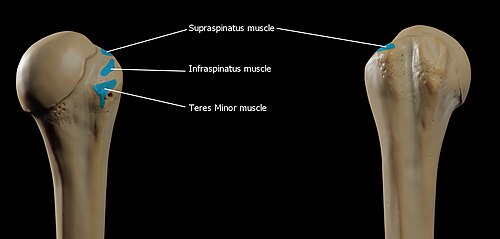
- Greater Tubercle: A large lateral projection where the supraspinatus, infraspinatus, and teres minor muscles attach. Its crest forms the lateral lip of the bicipital groove and provides an insertion point for the pectoralis major.
- Lesser Tubercle: A smaller, anterolateral projection that serves as the insertion point for the subscapularis muscle. Its crest forms the medial lip of the bicipital groove and hosts the teres major and latissimus dorsi muscles.
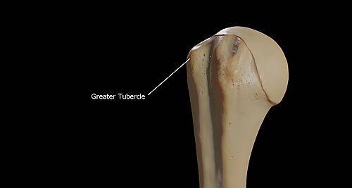
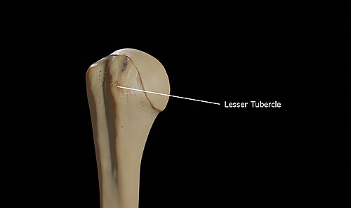
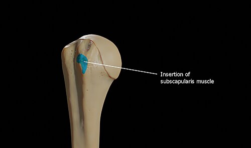
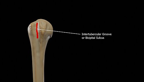
Separating these two tubercles is the bicipital groove (intertubercular groove). This deep channel lodges the long tendon of the biceps brachii muscle and transmits a branch of the anterior humeral circumflex artery.
The Shaft (Body of the Humerus)
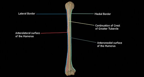
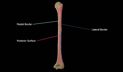
The shaft is a long, cylindrical section that transitions into a more prismatic shape as it descends. It is characterized by three surfaces and three borders.
Surfaces and Borders
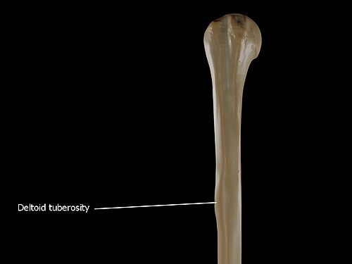
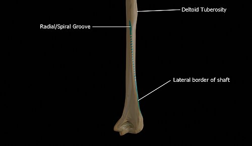
The anterolateral surface is smooth and rounded at the top, covered by the deltoid muscle. Mid-way down, it features the deltoid tuberosity, a rough elevation where the deltoid muscle inserts. The anteromedial surface is narrower and provides attachment for the coracobrachialis and brachialis muscles.
The posterior surface is largely covered by the two heads of the triceps brachii. A critical feature here is the radial sulcus (spiral groove), a shallow oblique depression that transmits the radial nerve and profunda artery.
Nutrient Foramen and Ridges
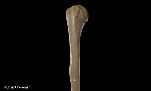
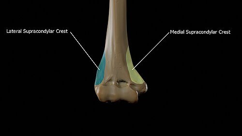
The nutrient foramen is located on the anteromedial surface, allowing nutrient arteries to enter the bone. As the shaft reaches the elbow, it forms the medial and lateral supracondylar ridges, which serve as origins for various forearm muscles, including the brachioradialis and pronator teres.
The Lower Extremity (Distal End)
The distal end of the humerus is flattened and curved, facilitating the complex movements of the elbow joint.
Articular Surfaces
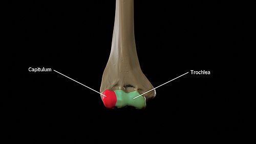
The articular surface is divided into two distinct parts:
- Capitulum: A smooth, rounded lateral eminence that articulates with the head of the radius.
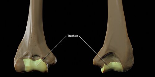
- Trochlea: A spool-shaped medial portion that fits into the semilunar notch of the ulna.
Fossae and Epicondyles
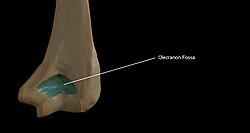
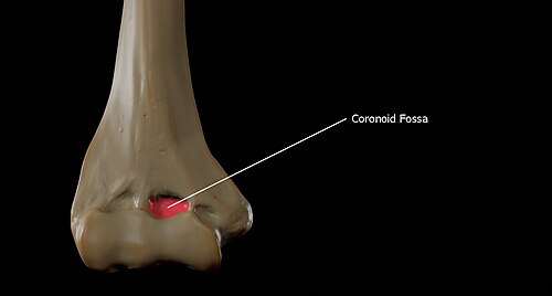
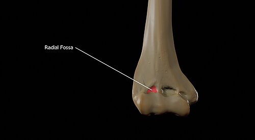
Three depressions, or fossae, allow for full joint range of motion: the coronoid fossa and radial fossa on the anterior side (used during flexion), and the olecranon fossa on the posterior side (used during extension).
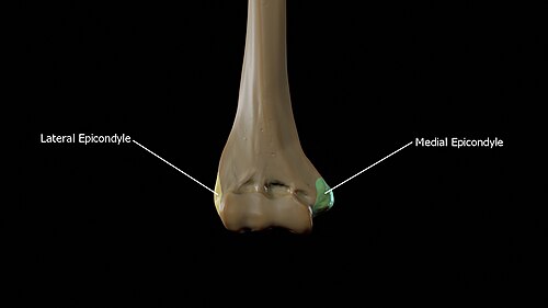
The distal end also features the medial and lateral epicondyles. The medial epicondyle is larger and more prominent, serving as the origin for forearm flexor muscles and providing a groove for the ulnar nerve.
Summary of Humeral Anatomy
| Region | Landmark | Primary Function/Association |
|---|---|---|
| Proximal | Surgical Neck | Common fracture site; Axillary nerve proximity |
| Proximal | Greater Tubercle | Attachment for Supraspinatus, Infraspinatus, Teres minor |
| Proximal | Lesser Tubercle | Attachment for Subscapularis |
| Shaft | Deltoid Tuberosity | Insertion point for Deltoid muscle |
| Shaft | Spiral Groove | Pathway for the Radial nerve |
| Distal | Capitulum | Articulation with the Radius |
| Distal | Trochlea | Articulation with the Ulna |
| Distal | Medial Epicondyle | Origin for forearm flexors; Ulnar nerve pathway |
Ossification and Development
The humerus is one of the first bones to ossify during embryonic development, starting in the shaft. Because this process is highly predictable, it is used as a biometric measurement to determine the gestational age of a fetus. At birth, only the shaft is ossified; the epiphyses (ends of the bone) remain cartilaginous. The medial head ossifies around 4 months, and the greater tuberosity around 10 months. Fusion typically begins at age 3 and is complete by age 13, though the growth plate persists until skeletal maturity around age 17.
Frequently Asked Questions
What is the difference between the anatomical neck and the surgical neck?
The anatomical neck is the groove immediately below the head where the joint capsule attaches. The surgical neck is a narrower area further down, below the tubercles, which is more prone to fractures.
Which nerve is most at risk during a mid-shaft humerus fracture?
The radial nerve is most at risk because it travels directly against the bone within the spiral groove (radial sulcus) at the mid-shaft.
What muscles attach to the greater tubercle?
The supraspinatus, infraspinatus, and teres minor muscles all attach to the greater tubercle.
How does the humerus articulate with the forearm?
The humerus articulates with the forearm at two points: the capitulum connects with the head of the radius, and the trochlea connects with the trochlear notch of the ulna.
Why is the humerus used in fetal biometrics?
Because the humerus is one of the first structures to ossify and does so at a predictable rate, its length can be measured via ultrasound to estimate the gestational age of a fetus.