Pituitary Gland: Anatomy, Hormones, and Endocrine Functions
The pituitary gland, also known as the hypophysis cerebri, is a vital endocrine gland found in vertebrates. In humans, this pea-sized organ is located at the base of the brain, protruding from the bottom of the hypothalamus. It sits securely within the sella turcica, a bony depression in the sphenoid bone known as the hypophyseal fossa.
Despite its small size—averaging about 1 cm in diameter and weighing between 0.5 and 1 gram—the pituitary gland acts as a master regulator. Together with the hypothalamus, it controls much of the body's endocrine system, influencing everything from growth and metabolism to reproduction and stress responses.
Key Facts
- Location: Base of the brain, below the hypothalamus in the sella turcica.
- Size: Approximately the size of a kidney bean (1 cm diameter).
- Structure: Divided into an anterior lobe (adenohypophysis) and a posterior lobe (neurohypophysis).
- Control: Both lobes are regulated by the hypothalamus.
- Primary Role: Secretes hormones that regulate growth, blood pressure, metabolism, and reproductive functions.
Anatomical Structure
The pituitary gland is composed of three distinct regions: the anterior lobe, the posterior lobe, and a small intermediate lobe that joins them. While the intermediate lobe is distinct in many animals (such as rats and mice), it is avascular and nearly absent in humans.
The Anterior Pituitary (Adenohypophysis)
The anterior lobe is the glandular portion of the organ. It originates from the oral ectoderm via an evagination called Rathke's pouch. This lobe is further divided into the pars tuberalis and the pars distalis, the latter making up roughly 80% of the gland. It contains specialized cells that synthesize and secrete a variety of essential hormones.
The Posterior Pituitary (Neurohypophysis)
Unlike the anterior lobe, the posterior pituitary is neural in composition and serves as an extension of the hypothalamus. It develops from the neuroectoderm of the floor of the third ventricle. It does not synthesize its own hormones; instead, it stores and releases neurohypophysial hormones produced by magnocellular neurosecretory cells in the hypothalamus.
Hormonal Functions and Regulation
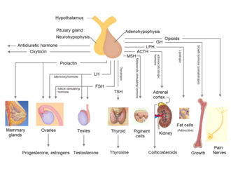
The pituitary gland regulates physiological processes by releasing specific hormones into the bloodstream. The anterior lobe is controlled by the hypothalamus via the hypothalamic-hypophysial portal system, a specialized capillary network.
Anterior Pituitary Hormones
| Hormone | Symbol | Target Organ | Primary Effect |
|---|---|---|---|
| Adrenocorticotropic hormone | ACTH | Adrenal gland | Secretion of glucocorticoids, mineralocorticoids, and androgens |
| Thyroid-stimulating hormone | TSH | Thyroid gland | Secretion of thyroid hormones |
| Follicle-stimulating hormone | FSH | Gonads | Growth of reproductive system |
| Luteinizing hormone | LH | Gonads | Sex hormone production |
| Growth hormone | GH | Liver, adipose tissue | Promotes growth; lipid and carbohydrate metabolism |
| Prolactin | PRL | Mammary glands, ovaries, testes | Lactation and sex hormone regulation |
Posterior Pituitary Hormones
The posterior lobe stores and secretes two primary hormones synthesized in the hypothalamus:
- Vasopressin (ADH): Regulates hydroelectrolytic stability and water reabsorption in the kidneys.
- Oxytocin: Stimulates uterine contractions during labor and facilitates human attachment. Oxytocin is notable for creating a positive feedback loop during childbirth.
Development and Growth
The development of the pituitary gland occurs in stages during embryonic growth. In weeks 5 and 6, Rathke's pouch grows toward the brain and eventually detaches from the oral cavity to form the adenohypophysis. Simultaneously, between weeks 4 and 8, the infundibulum elongates downward from the diencephalon to form the posterior pituitary.
By weeks 12 to 16 of gestation, the anterior pituitary begins producing critical hormones, such as growth hormone (GH) and ACTH, which are essential for fetal development.
Clinical Significance
Because the pituitary gland controls so many systems, any imbalance in hormone production can lead to significant health issues. These are generally categorized as hyperpituitarism (over-secretion) or hypopituitarism (under-secretion).
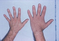
- Gigantism and Acromegaly: Caused by an excess of growth hormone. Gigantism occurs in childhood, while acromegaly occurs in adults.
- Central Diabetes Insipidus: Resulting from a deficiency of vasopressin.
- Hypothyroidism: Often caused by a deficiency in thyroid-stimulating hormone (TSH).
- Pituitary Adenomas: Mostly benign tumors that can disrupt normal hormone secretion.
Frequently Asked Questions
What is the difference between the anterior and posterior pituitary?
The anterior pituitary is a true glandular structure that synthesizes its own hormones, originating from oral ectoderm. The posterior pituitary is an extension of the brain (neural tissue) that stores and releases hormones produced by the hypothalamus.
How does the hypothalamus control the pituitary gland?
The hypothalamus controls the anterior pituitary via the hypothalamic-hypophysial portal system (blood vessels) and controls the posterior pituitary through direct neural projections (axons) that travel down the pituitary stalk.
What happens if the pituitary gland produces too much growth hormone?
Excess growth hormone in children leads to gigantism, characterized by excessive height. In adults, it causes acromegaly, which results in the enlargement of bones in the hands, feet, and face.
What is Rathke's pouch?
Rathke's pouch is an evagination of the oral ectoderm that develops during the fifth and sixth weeks of gestation, eventually forming the anterior lobe of the pituitary gland.
Which hormone is responsible for water balance in the body?
Vasopressin, also known as antidiuretic hormone (ADH), is stored in the posterior pituitary and regulates water balance by controlling how much water the kidneys reabsorb.