Thyroid Gland: Anatomy, Hormone Synthesis, and Clinical Significance
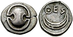
The thyroid gland is a vital component of the endocrine system, responsible for regulating the body's metabolic rate, cardiovascular function, and developmental growth. Named after the Greek word for "shield" due to its distinct shape, this gland acts as a chemical control center, secreting hormones that influence nearly every cell in the human body.
From its embryonic origins to its complex microanatomy, the thyroid operates through a sophisticated feedback loop involving the hypothalamus and the pituitary gland to maintain internal homeostasis.
Key Facts
- Location: Surrounds the cricoid and tracheal cartilages in the neck.
- Primary Hormones: Produces thyroxine (T4) and triiodothyronine (T3).
- Essential Nutrient: Iodine is critical for the synthesis of thyroid hormones.
- Regulation: Controlled by Thyroid Stimulating Hormone (TSH) and Thyrotropin Releasing Hormone (TRH).
- Development: TSH becomes measurable at 11 weeks of fetal development.
Anatomy and Structure
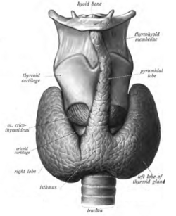
The thyroid gland typically consists of two lobes that wrap around the trachea. While the standard structure is well-defined, anatomical variations occur, such as the presence of a pyramidal lobe emerging from the center of the gland.
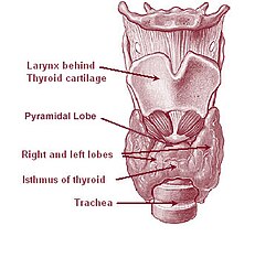
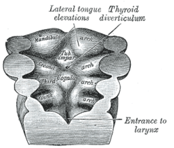
The gland is supplied by the superior and inferior thyroid arteries and drained by the superior, middle, and inferior thyroid veins. Its development begins as a thyroid diverticulum, an extension of endoderm into the second pharyngeal arch.
Microanatomy
Under a microscope, the thyroid is organized into spherical structures called follicles. Each follicle consists of a layer of follicular cells surrounding a central lumen filled with colloid, a protein-rich fluid where hormone precursors are stored.
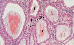
In addition to follicular cells, the gland contains parafollicular cells (C-cells), which are responsible for the synthesis and secretion of calcitonin, a hormone involved in calcium regulation.
Hormone Synthesis and Function
The production of thyroid hormones is a complex biochemical process that relies heavily on the active transport of iodide from the blood into the follicular cells.
The Synthesis Process
- Thyroglobulin Production: Follicular cells synthesize thyroglobulin, which is secreted into the colloid via exocytosis.
- Iodide Transport: A sodium-iodide (Na/I) symporter pumps iodide into the cell, and the transporter pendrin moves it into the lumen.
- Oxidation and Iodination: The enzyme thyroid peroxidase oxidizes iodide to iodine, which then attaches to tyrosyl residues on the thyroglobulin chain.
- Coupling and Release: Adjacent tyrosyl residues pair together. The complex is re-absorbed into the cell via endocytosis, where proteases liberate T3 and T4 into the bloodstream.
![Synthesis of the thyroid hormones, as seen on an individual thyroid follicular cell:[33] - Thyroglobulin is synthesized in the rough endoplasmic reticulum and follows the secretory pathway to enter the colloid in the lumen of the thyroid follicle by exocytosis. - Meanwhile, a sodium-iodide (Na/I) symporter pumps iodide (I−) actively into the cell, which previously has crossed the endothelium by largely unknown mechanisms. - This iodide enters the follicular lumen from the cytoplasm by the transporter pendrin, in a purportedly passive manner. - In the colloid, iodide (I−) is oxidized to iodine (I0) by an enzyme called thyroid peroxidase. - Iodine (I0) is very reactive and iodinates the thyroglobulin at tyrosyl residues in its protein chain (in total containing approximately 120 tyrosyl residues). - In conjugation, adjacent tyrosyl residues are paired together. - The entire complex re-enters the follicular cell by endocytosis. - Proteolysis by various proteases liberates thyroxine and triiodothyronine molecules, which enters the blood by largely unknown mechanisms.](/images/50/c8/50c8118aa613b6c9249bcf27e5c99aabb1a8a991b1b8992a7d4e91000c717ab2.png)
Hormonal Activity and Transport
The gland primarily secretes thyroxine (T4), comprising 80–90% of its output, and triiodothyronine (T3), comprising 10–20%. In the blood, most of these hormones are bound to proteins: 70% to thyroxine-binding globulin, 15% to albumin, and 10% to transthyretin. Only the free fractions (0.03% for T4 and 0.3% for T3) are biologically active.
Notably, up to 85% of circulating T3 is produced in peripheral organs through the conversion of T4 by enzymes called iodothyronine deiodinases.
Regulation and Development
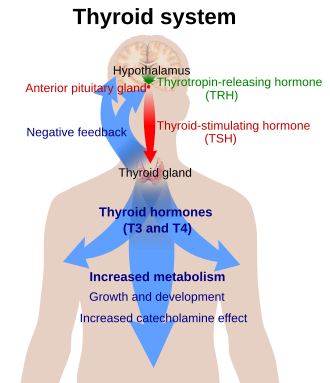
The thyroid is governed by a negative feedback loop. The hypothalamus releases Thyrotropin Releasing Hormone (TRH), which stimulates the pituitary to release Thyroid Stimulating Hormone (TSH). TSH then triggers the thyroid to produce T3 and T4. As levels of these hormones rise, they inhibit the further release of TRH and TSH.
Fetal Development
Thyroid function is critical during pregnancy. While TSH is measurable at 11 weeks, T4 reaches self-sufficient levels by 18–20 weeks. Sufficient iodine is mandatory during this period to prevent neurodevelopmental disorders.
Clinical Significance
Dysfunction of the thyroid can lead to various metabolic and physical disorders. Hyperthyroidism occurs when the gland is overactive, while hypothyroidism occurs when it is underactive.
Common Conditions
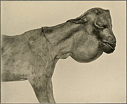
- Goitre: An abnormal enlargement of the thyroid gland, often caused by iodine deficiency.
- Graves' Disease: An autoimmune disorder leading to hyperthyroidism.
- Hashimoto's Thyroiditis: An autoimmune condition causing hypothyroidism.
- Congenital Iodine Deficiency Syndrome: A severe condition resulting from a lack of iodine during development.
![Child affected by Congenital iodine deficiency syndrome, associated with a lack of iodine.[75]](/images/d1/25/d12598ea5f8ff0c0801bb9341dddfbde8ce23b68397961f96d98604738ae4339.jpg)
Summary of Thyroid Components
| Feature | Details |
|---|---|
| Primary Hormones | T3 (Triiodothyronine), T4 (Thyroxine) |
| Regulatory Hormones | TRH (Hypothalamus), TSH (Pituitary) |
| Key Enzyme | Thyroid Peroxidase (TPO) |
| Essential Element | Iodine |
| C-Cell Product | Calcitonin |
Frequently Asked Questions
What is the difference between T3 and T4?
T4 (thyroxine) is produced in larger quantities by the thyroid gland and acts as a prohormone. T3 (triiodothyronine) is the more biologically active form; most of it is created when T4 is converted by deiodinase enzymes in peripheral tissues.
Why is iodine important for the thyroid?
Iodine is a central ingredient in the synthesis of thyroid hormones. Without sufficient iodine, the gland cannot produce T3 and T4, which can lead to the development of a goitre or congenital developmental issues.
What is the role of the pituitary gland in thyroid function?
The pituitary gland secretes Thyroid Stimulating Hormone (TSH), which tells the thyroid gland to absorb iodide and produce T3 and T4. This process is regulated by negative feedback from the hormones themselves.
What are parafollicular cells?
Also known as C-cells, these are specialized cells located between the thyroid follicles. Unlike follicular cells, they do not produce T3 or T4; instead, they synthesize and secrete calcitonin.
How does the thyroid affect fetal development?
Thyroid hormones are essential for healthy neurodevelopment. The fetus becomes self-sufficient in T4 around 18–20 weeks, and adequate iodine levels are required to prevent permanent cognitive and developmental impairments.