Endostyle: The Evolutionary Precursor to the Thyroid Gland
The endostyle is a specialized organ found in invertebrate chordates, including tunicates and lancelets, as well as in the larval stage of vertebrate lampreys. Serving as a critical component of the filter-feeding mechanism, this organ provides a fascinating glimpse into evolutionary history, as it is the biological precursor to the thyroid gland found in higher vertebrates.
Because the endostyle appears across all three chordate lineages, scientists believe it originated in a common ancestor. This development coincided with a shift toward internal feeding, allowing these organisms to extract suspended food particles from the surrounding water more efficiently.
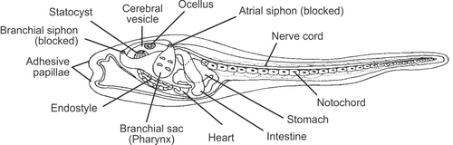
How the Endostyle Works
The primary function of the endostyle is to facilitate feeding through a sophisticated filtration process. The organ produces a sticky mucus that captures food particles suspended in the water. Once the particles adhere to this mucus, the filtered water is expelled through the gill slits. Meanwhile, the combined food and mucus are swept into the esophagus by the coordinated movements of cilia (tiny hair-like projections) that coat the organ.
The Evolutionary Link to the Thyroid
One of the most significant aspects of the endostyle is its homology—a shared evolutionary origin—with the thyroid gland. In larval lampreys, known as ammocoetes, the endostyle undergoes metamorphosis to become the thyroid gland in adult lampreys.
This connection was first highlighted by the organ's ability to concentrate iodine, a primary function of the modern thyroid. While early hypotheses in 1873 suggested the ammocoete endostyle functioned similarly to the tunicate hypobranchial groove, later research shifted toward its chemical properties. By 1963, studies confirmed that both cephalochordate and tunicate endostyles could capture iodine. Modern genetic research has further supported this link, showing that the development of the endostyle involves transcription factors similar to those that govern thyroid development in vertebrates.
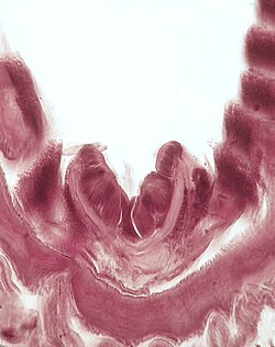
Key Facts
- Function: Primarily used for filter-feeding by producing mucus to trap food particles.
- Evolution: Evolved into the thyroid gland in vertebrate chordates.
- Distribution: Found in tunicates, lancelets, and larval lampreys (ammocoetes).
- Chemical Property: Capable of concentrating iodine, mirroring thyroid activity.
- Mechanism: Uses cilia to move trapped food toward the esophagus.
Detailed Anatomy and Zonal Structure
When viewed in a transverse section, the endostyle is organized into several distinct zones, each with unique cellular characteristics and functions:
| Zone | Key Characteristics | Primary Features/Contents |
|---|---|---|
| Zone 1 | Bottom portion | Tall cilia, glycogen, acid mucopolysaccharides |
| Zone 2 | Ventral glandular tract | Acid mucous material; high secretory activity |
| Zone 3 | Ciliated bands | Granulated apical border |
| Zone 4 | Dorsal-like glandular tract | Rough granulation; pyroninophilic material |
| Zone 5 | Granulated surface | PA/S-positive droplets; potential iodination center |
| Zone 6 | Lateral edge | Small cytoplasmic granulation |
| Zone 7 | Iodine trap | Various granule sizes; no secretory activity |
| Zone 8 | Lateral edge | High density of cilia; acidic mucosal material |
| Zone 11 | Lateral granulations | Unique cytoplasm; no cilia |
Cellular Makeup
The endostyle consists of different cell types that manage its structural and secretory needs:
- Type 1 Cells: These form the gland cylinders. They feature enlarged circular nuclei and a large nucleolus. Ventral cylinders show a strong cyanophile reaction, while dorsal cylinders show a weak PA/S-positive reaction.
- Type 2 Cells: These are cylinder opening cells, subdivided into 2a, 2b, and 2c. Type 2c cells are particularly easy to recognize due to their rich granulation.
Secretory Activity and Iodine Metabolism
Secretory activity is primarily handled by the gland cells, which synthesize proteins rich in -SS and -SH bridges and release disease-resistant carbohydrates. The ventral glandular region secretes a mix of proteins and mucopolysaccharides (including mucoproteins or glycoproteins), while the dorsal glandular region is rich in lipids, cystin, or cysteine.
Crucially, these glandular regions are not involved in iodine metabolism. Instead, the ability to trap radioiodine is linked to specific zones. While some research points to Zone 5, others suggest Zone 7 or Zone 8 as the primary iodination centers. This capacity for iodine collection and breakdown is what establishes the endostyle as the evolutionary precursor to the thyroid.
Frequently Asked Questions
What is the primary purpose of the endostyle?
The endostyle is primarily used for filter-feeding. It secretes mucus to trap food particles from the water, which are then transported to the esophagus by cilia.
How does the endostyle relate to the human thyroid?
The endostyle is homologous to the thyroid gland. It shares a common evolutionary origin and possesses the same ability to concentrate iodine, which is a hallmark of thyroid function.
In which animals can the endostyle be found?
It is found in invertebrate chordates such as tunicates and lancelets, as well as in the larval stage (ammocoetes) of lampreys.
Which zones of the endostyle are responsible for secretion?
Secretory activity occurs mainly in the ventral and dorsal glandular regions, specifically within Zone 2 and Zone 4, with some accumulation also occurring in Zone 5.
Why do different studies disagree on the iodination center of the endostyle?
Discrepancies may arise because some zones may simply trap iodine due to the presence of cilia, while other zones are responsible for the actual breakdown and metabolism of iodine materials.