Coliform Bacteria: Characteristics, Identification, and Public Health Impact
Coliform bacteria are a diverse group of Gram-negative bacilli that play a critical role in both natural ecosystems and human health. While many of these organisms are harmless commensals—bacteria that live in a symbiotic relationship with their host—certain strains can become opportunistic pathogens. Because many coliforms are almost exclusively of fecal origin, they serve as essential biological indicators for fecal contamination in water and food supplies.
Key Facts
- E. coli is the most common coliform in the human intestine and a primary indicator of fecal contamination.
- Citrobacter infections carry a significant mortality rate between 33% and 48%, particularly in infants and immunocompromised patients.
- Klebsiella species are distinguished by a protective polysaccharide capsule that allows them to survive drying for several months.
- EMB Agar is a primary tool for differentiation, where E. coli produces a characteristic metallic green sheen.
- Detection methods range from traditional culture media like Violet Red Bile Agar to advanced molecular techniques like PCR and chemiluminescent in-situ hybridization.
Common Genera of Coliform Bacteria
Escherichia
Escherichia species are the most frequent cause of coliform-related diseases in humans. Escherichia coli (E. coli) is the most prevalent organism in the human intestine. While most strains are harmless, some acquire virulence through horizontal gene transfer—the movement of genetic material between unicellular and multicellular organisms.
Pathogenic E. coli are categorized into several pathotypes based on the gastrointestinal syndromes they cause: diarrheagenic (DEC), enterotoxigenic (ETEC), enteroaggregative (EAEC), enteroinvasive (EIEC), enteropathogenic (EPEC), and Shiga toxin–producing (STEC), which includes enterohemorrhagic (EHEC) strains. Symptoms of infection can include vomiting, stomach cramps, fever, and bloody diarrhea, though they can also cause respiratory illnesses and pneumonia.
Enterobacter
Enterobacter are motile, flagellated bacilli. They are known to cause a variety of infections, including respiratory tract infections, bacteremia (bacteria in the blood), urinary tract infections, and surgical site infections. In severe cases, they may lead to osteomyelitis (bone infection), sinusitis, or meningitis.
Klebsiella
Klebsiella are non-motile bacilli typically measuring 1–2 μm in length. They are facultative anaerobes, meaning they can grow with or without oxygen. Klebsiella pneumoniae is the most common species found in humans, animals, soil, and sewage. These bacteria produce a mucosal outer surface and a complex acid polysaccharide capsule that provides significant environmental resilience.
Citrobacter
Citrobacter are peritrichous (having flagella all over the cell surface) facultative anaerobic bacilli. While they typically inhabit the intestinal flora without causing harm, they can cause severe infections if they enter other parts of the body, leading to brain abscesses, pneumonia, joint infections, meningitis, and intra-abdominal sepsis.
Laboratory Identification and Differentiation
Distinguishing between these genera requires specific culture media and biochemical tests. For example, Enterobacter is differentiated from Escherichia using an indole test; Enterobacter is indole negative, while Escherichia is positive. Enterobacter is further distinguished from Klebsiella by its motility.
Eosin Methylene Blue (EMB) agar is a vital tool for identifying coliforms. This medium inhibits Gram-positive bacteria and reacts differently based on the organism's ability to ferment lactose. Strong lactose fermenters appear dark blue, purple, or black. E. coli, a strong fermenter, produces a distinct metallic green sheen on the dark purple medium.
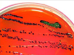
Other identification methods include Tryptone Bile X-Glucuronide (TBX), where E. coli appears as blue or green colonies after 24 hours of incubation at an optimal temperature of 37 °C.
Summary of Coliform Genera
| Genus | Motility | Key Characteristics | Common Associated Infections |
|---|---|---|---|
| Escherichia | Mostly Motile | Metallic green on EMB; lactose fermenter | UTIs, bloody diarrhea, pneumonia |
| Enterobacter | Motile | Indole negative; lactose fermenter | Bacteremia, respiratory infections, osteomyelitis |
| Klebsiella | Non-motile | Polysaccharide capsule; mucosal colonies | Pneumonia, UTIs |
| Citrobacter | Peritrichous | Facultative anaerobic bacilli | Brain abscesses, sepsis, joint infections |
Outbreaks and Public Health
Contaminated food sources frequently lead to coliform outbreaks. In late 2021, an investigation by the FDA and the Minnesota Department of Agriculture linked ten cases of E. coli O157:H7 across seven US states to contaminated spinach. This was confirmed using whole genome sequencing to match the strain in the spinach to the strain in the patients.
Similarly, between December 2021 and January 2022, Canada reported fourteen cases of E. coli O157. The Public Health Agency of Canada (PHAC) and other agencies traced the source to a specific brand of Original Kimchi, leading to a recall of Hankook Original Kimchi in early 2022.
Detection Methods in Drinking Water
Molecular Techniques
- PCR (Polymerase Chain Reaction): This method amplifies the beta-galactosidase gene to detect general coliforms. To specifically identify E. coli, the beta-D glucuronidase gene or verotoxin genes are amplified.
- Chemiluminescent in-situ hybridization: This uses soybean peroxidase-labeled peptide nucleic acid (PNA) probes that bind to the 16S rRNA of E. coli. When a substrate is added, light is produced, marking the location of each colony.
Culture-Based Methods
- Violet Red Bile Agar: Lactose-fermenting coliforms produce pink colonies. These are confirmed by transferring them to brilliant green lactose bile (BGLB) medium; gas production confirms the presence of coliforms.
- Membrane Filter Method: Samples are filtered and placed on M-endo or LES Endo Agar. Pinkish-red colonies with a green metallic sheen are identified and then confirmed via lauryl tryptose (LST) and BGLB inoculation for gas production.
Frequently Asked Questions
How is E. coli distinguished from other coliforms on EMB agar?
While many coliforms appear dark blue or purple due to lactose fermentation, E. coli produces a characteristic metallic green sheen on the dark purple medium.
What is the significance of the polysaccharide capsule in Klebsiella?
The capsule, composed of complex acid polysaccharides, allows Klebsiella to withstand drying for several months, increasing its environmental persistence.
Why is E. coli used as an indicator of fecal contamination?
Unlike the general coliform group, E. coli are almost exclusively of fecal origin, making their presence a reliable confirmation that fecal contamination has occurred.
What is the difference between Enterobacter and Klebsiella in the lab?
The primary difference used for laboratory distinction is motility; Enterobacter are motile, whereas Klebsiella are non-motile.
How does PCR detect different types of coliforms?
PCR targets specific genes: the beta-galactosidase gene is used for general coliforms, while the beta-D glucuronidase or verotoxin genes are targeted to identify E. coli and verotoxin-producing strains, respectively.