Cerebral Hemispheres
The cerebrum is the largest portion of the vertebrate brain, characterized by its division into two distinct cerebral hemispheres. While these two halves appear as macroscopic mirror images, they work in tandem to process sensory information, execute motor commands, and facilitate complex cognitive functions. The two sides are separated by a deep groove called the longitudinal fissure, yet they remain deeply interconnected to ensure the brain operates as a unified system.
The primary bridge between these hemispheres is the corpus callosum, a massive bundle of nerve fibers that integrates sensory and motor signals. While other smaller fiber bundles exist—such as the anterior commissure, posterior commissure, and the fornix—the corpus callosum is the dominant structure for inter-hemispheric communication in placental mammals.
Key Facts
- Composition: Consists of an outer layer of gray matter (cerebral cortex) and an inner core of white matter.
- Division: Split into left and right hemispheres by the longitudinal fissure.
- Lobes: Each hemisphere is divided into frontal, parietal, occipital, and temporal lobes.
- Connectivity: The corpus callosum is the primary neural bridge between the two sides.
- Development: Derived from the telencephalon, appearing five weeks after conception.
Anatomical Structure and Composition
The cerebral hemispheres are composed of two primary tissue types. The thin outer layer, known as the cerebral cortex (from the Latin word for "bark of a tree"), is made of gray matter. This tissue contains neuronal cell bodies, dendrites, and synapses. Beneath the cortex lies a larger volume of white matter, which consists of axons and myelin.
Deep within the hemispheres, the white matter forms a core known as the centrum semiovale. This area is supplied by the superficial middle cerebral artery. Other internal structures include the lateral ventricles and the basal ganglia.
Each hemisphere is further organized into four lobes: the frontal, parietal, occipital, and temporal lobes. A prominent fissure called the central sulcus serves as the boundary between the frontal and parietal lobes, as well as the dividing line between the primary motor cortex and the primary somatosensory cortex.
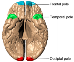
Three of these lobes feature distinct "poles":
- Frontal pole: The rounded frontmost part of the frontal lobe.
- Occipital pole: The more pointed posterior end of the occipital lobe.
- Temporal pole: Located in the anterior part of the middle cranial fossa.
Detailed Internal Anatomy
When the upper portion of a hemisphere is removed (approximately 1.25 cm above the corpus callosum), the oval-shaped centrum semiovale is revealed. This white matter is surrounded by a convoluted margin of gray substance and marked by puncta vasculosa—minute red dots caused by blood escaping from divided vessels.
The margins of the hemispheres that overlap the corpus callosum are termed the labia cerebri, which are part of the cingulate gyrus. The groove separating the labium from the upper surface of the corpus callosum is known as the callosal sulcus.
Development and Growth
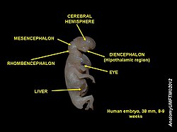
The cerebral hemispheres originate from the telencephalon. They first appear five weeks after conception as bilateral invaginations of the walls. During development, the hemispheres grow in a C-shape and then curve back, pulling internal structures like the ventricles along with them.
Communication with the lateral ventricles is facilitated by the intraventricular foramina (foramina of Monro). Additionally, the choroid plexus develops from vascular mesenchyme and ependymal cells.
Hemisphere Lateralization and Function
While popular psychology often suggests a strict divide between "logical" left-brain and "creative" right-brain thinking, scientific evidence shows that most higher-level functions are distributed across both hemispheres. Lateralization—the localization of function in one hemisphere—is more common in low-level perceptual functions.
A primary example of lateralization is found in language processing. Broca's and Wernicke's areas are frequently located exclusively in the left hemisphere, often corresponding to the hemisphere opposite the individual's dominant hand. Generally, linear reasoning (grammar and word production) tends to be left-lateralized, while holistic reasoning (intonation and emphasis) is often right-lateralized.
Perceptual and motor signals are contralateral, meaning information from one side of the body is processed by the opposite hemisphere, and motor signals are sent from the opposite side to the body.
Asymmetries Between Hemispheres
Despite their similar appearance, the hemispheres exhibit microscopic and physical asymmetries:
- Size: The right hemisphere is slightly larger.
- Chemistry: The right side has higher levels of norepinephrine, while the left has higher levels of dopamine.
- Sensitivity: The right hemisphere is more sensitive to testosterone.
- Tissue Distribution: The right side contains more white matter (longer axons), while the left contains more gray matter (cell bodies).
Summary of Cerebral Hemisphere Characteristics
| Feature | Left Hemisphere | Right Hemisphere |
|---|---|---|
| Dominant Neurotransmitter | Dopamine | Norepinephrine |
| Tissue Prevalence | More Gray Matter | More White Matter |
| Language Function | Linear (Grammar, Word Production) | Holistic (Intonation, Emphasis) |
| Physical Size | Slightly Smaller | Slightly Larger |
| Hormonal Sensitivity | Standard | Higher sensitivity to Testosterone |
Clinical Significance
Medical conditions and interventions can specifically target the structures of the cerebral hemispheres. Infarcts (tissue death due to lack of blood supply) can occur within the centrum ovale.
In severe cases of epilepsy, surgeons may perform a corpus callosotomy, severing the corpus callosum to stop the spread of seizures between hemispheres. In extreme cases where seizures are unresponsive to other treatments, a hemispherectomy—the removal or disabling of an entire hemisphere—may be performed.
Frequently Asked Questions
What is the main difference between gray and white matter?
Gray matter is the outer layer (cerebral cortex) consisting of neuronal cell bodies, dendrites, and synapses. White matter is the inner layer consisting of axons and myelin, which facilitate communication between different brain regions.
Does the "left-brain vs. right-brain" personality theory hold true?
No. While some low-level functions and specific language areas are lateralized, most complex cognitive functions are distributed across both hemispheres rather than being confined to one side.
What is the role of the corpus callosum?
The corpus callosum is a large bundle of nerve fibers that connects the left and right cerebral hemispheres, allowing them to integrate sensory and motor signals.
How do the hemispheres control the body?
The brain uses contralateral control, meaning the left hemisphere processes sensory information and controls motor movements for the right side of the body, and vice versa.
What are the poles of the cerebrum?
The poles are the extreme ends of the lobes: the frontal pole (front of the frontal lobe), the occipital pole (back of the occipital lobe), and the temporal pole (front of the temporal lobe).