Organogenesis: How Embryonic Germ Layers Form Organs
Organogenesis is the critical phase of embryonic development that begins immediately following gastrulation and continues until birth. During this stage, the three primary germ layers—the ectoderm, mesoderm, and endoderm—transform into the complex tissues and internal organs that sustain an organism's life.
This transformation is driven by differentiation, a process where less-specialized cells become highly specialized by expressing specific sets of genes. This process is governed by cell signaling cascades, which ensure that organs develop at the correct time and in the precise locations required for survival. These signals include juxtacrine signaling (between adjacent cells), paracrine signaling (between neighboring cells over short distances), and autocrine signaling (where a cell produces and receives its own signal).
Because studying human embryos presents ethical and practical challenges, scientists often use model organisms such as zebrafish, chickens, mice, and Xenopus. Additionally, researchers utilize organoids—simplified, in vitro organ structures grown from adult, embryonic, or induced pluripotent stem cells—to study human development without the use of an embryo.
Key Facts
- Organogenesis follows gastrulation and lasts until birth.
- The three germ layers (ectoderm, mesoderm, endoderm) are the blueprints for all body tissues.
- In humans, internal organs typically begin developing between 3 and 8 weeks after fertilization.
- Neurulation is the specific process that forms the neural tube, the precursor to the central nervous system.
- Unlike animals, plant organogenesis occurs continuously throughout the plant's life.
The Three Germ Layers and Their Derivatives
Each germ layer is responsible for producing specific sets of tissues and organs. The interaction between these layers and their surrounding environment determines the final structure of the organism.
- Endoderm: The innermost layer, which forms the epithelial linings of the respiratory and gastrointestinal systems, as well as the liver, lungs, and pancreas.
- Mesoderm: The middle layer, which gives rise to the heart, blood, kidneys, muscles, and connective tissues.
- Ectoderm: The outermost layer, which develops into the epidermis of the skin, the brain, and the rest of the nervous system.
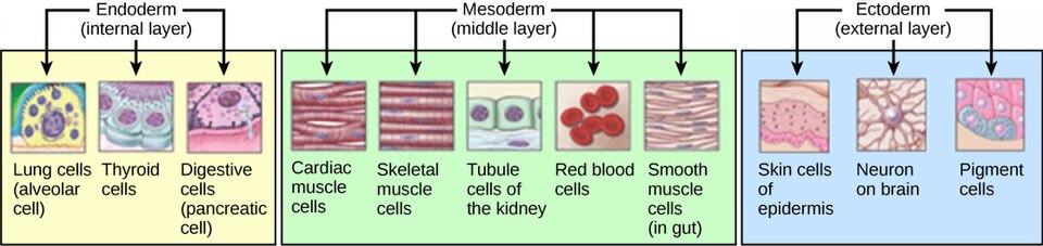
| Germ Layer | Position | Primary Organs/Tissues Produced |
|---|---|---|
| Ectoderm | Outermost | Epidermis, brain, nervous system, melanocytes |
| Mesoderm | Middle | Heart, blood, kidneys, skeletal/smooth/cardiac muscle |
| Endoderm | Innermost | Lungs, liver, pancreas, gastrointestinal lining |
Mechanisms of Organ Formation
The scientific understanding of organogenesis was shaped by early 19th-century pioneers. Caspar Friedrich Wolff proposed that organs form through the rhythmic folding and arrest of membranes. Heinz Christian Pander later identified the three germ layers and noted that their differentiation is influenced by surrounding tissues. Karl Ernst von Baer further refined these ideas, establishing laws of embryology stating that embryos of different species appear similar early on and move from generalized to specified features as they develop.
In humans, organs are formed through three primary physical processes:
- Folds: The germinal sheet of cells folds to create enclosed tubes.
- Splits: Pockets form in the cell sheet, creating vesicles or elongations (common in glands and lungs).
- Condensation: Cells cluster together to form denser tissue structures.
Neurulation and the Central Nervous System
For chordates, a pivotal event is the development of the notochord, which induces the formation of the neural plate. This leads to neurulation—the process of forming the neural tube from the ectoderm. In humans, this occurs around the third week of gestation.
Neurulation occurs in three distinct modes:
- Primary Neurulation: The edges of the neural plate rise to form neural folds and a groove, which then fuse to create the anterior portion of the neural tube.
- Secondary Neurulation: Mesenchymal cells (mobile, non-epithelial cells) migrate under the epidermis to form the posterior end of the tube.
- Junctional Neurulation: A transition zone combining both folding and mesenchymal migration.
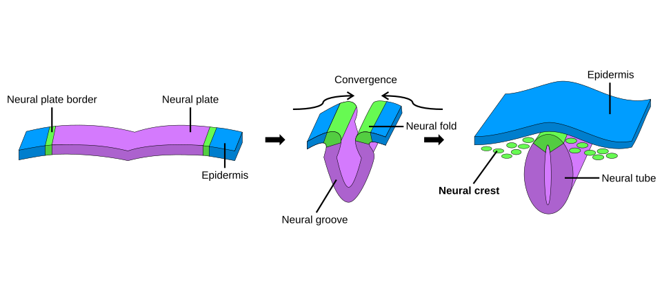
Once the neural tube closes and separates from the epidermis, it develops into the brain and spinal cord. Failure in this closure can lead to neural tube defects, such as spina bifida or anencephaly. Additionally, neural crest cells migrate from the tube to form various structures, including bones, muscles, and components of the central nervous system.
Organogenesis in Plants
Plant organogenesis differs fundamentally from animal development because it is a continuous process. In the shoot, shoot apical meristems constantly produce new lateral branches, leaves, flowers, or fruits. In the root, new lateral roots emerge from internal tissues, such as the xylem-pole pericycle in Arabidopsis thaliana.
Plants also possess a unique ability to de-differentiate. In the presence of specific hormones—primarily auxins and cytokinins—most plant tissues can revert to a mass of totipotent stem cells called a callus. Depending on the hormone concentrations in the medium, these cells can then undergo organogenesis to regenerate entire plants in tissue culture.
Frequently Asked Questions
What is the difference between gastrulation and organogenesis?
Gastrulation is the earlier phase that establishes the three primary germ layers (ectoderm, mesoderm, and endoderm). Organogenesis is the subsequent phase where these layers differentiate into specific organs and tissues.
What happens if the neural tube fails to close?
Failure of the neural tube to close properly can result in serious developmental conditions known as neural tube defects, most notably spina bifida and anencephaly.
How do organoids help in medical research?
Organoids are simplified versions of organs grown in vitro from stem cells. They allow scientists to study human organ development and disease without the ethical concerns or practical difficulties associated with studying human embryos.
How does plant organogenesis differ from animal organogenesis?
While animal organogenesis primarily occurs during embryonic development, plant organogenesis happens continuously throughout the plant's life. Additionally, plants can de-differentiate their cells into a callus to regenerate organs.
What are the three types of cell signaling involved in differentiation?
Differentiation is driven by juxtacrine signaling (between touching cells), paracrine signaling (between nearby cells), and autocrine signaling (a cell signaling to itself).