Neurocranium: Anatomy and Evolution of the Braincase
The neurocranium, commonly referred to as the braincase, brainpan, or brainbox, is the specialized upper and posterior portion of the skull. Its primary biological function is to provide a rigid, protective enclosure for the brain. In humans, this structure includes the calvaria (the skullcap), while the remaining portion of the skull is categorized as the facial skeleton.
In the field of comparative anatomy, the term neurocranium is often used interchangeably with the endocranium or the chondrocranium, depending on the species and the developmental stage being discussed.
Key Facts
- The human neurocranium consists of eight primary bones.
- It is divided into a membranous part (flat bones) and a cartilaginous part (the chondrocranium).
- The foramen magnum is the critical opening that allows the neural spine to exit the skull.
- Evolutionarily, the human neurocranium has expanded to overgrow the splanchnocranium (facial skeleton) to accommodate a larger brain.
- Development begins with paraxial mesoderm and ectomesenchyme.
Human Neurocranium Structure
The human braincase is a complex assembly of bones that protect the central nervous system. It is structurally divided into two distinct portions: the membranous part, composed of flat bones that wrap around the brain, and the cartilaginous part, known as the chondrocranium, which forms the base of the skull.
The human neurocranium is typically composed of the following eight bones:
- Frontal bone (1)
- Parietal bones (2)
- Temporal bones (2)
- Occipital bone (1)
- Sphenoid bone (1)
- Ethmoid bone (1)
While the auditory ossicles (the three tiny bones in each middle ear) are located within this region, they are generally not classified as part of the neurocranium. Additionally, some individuals may possess extra sutural bones.
Below the main structure lies a network of foramina (openings) and specialized bones. The most prominent is the foramen magnum, which houses the neural spine. In this same region, the auditory bullae assist in the process of hearing.
Development and Biological Variation
The neurocranium originates from paraxial mesoderm, with additional contributions from ectomesenchyme. In certain vertebrates, such as Chondrichthyes (cartilaginous fish), this portion of the cranium does not undergo ossification, meaning it is not replaced by bone through endochondral ossification.
Among mammals, the size and shape of the neurocranium vary significantly. Some species exhibit specific ridges, such as temporal crests, on the roof of the skull.
Comparative Anatomy in Other Vertebrates
Across different species, the neurocranium is formed by the combination of the endocranium, the skull roof, and the lower portions of the cranial vault. In humans, the neurocranium has evolved to cover both the back and the top of the skull, eventually overgrowing the facial skeleton. This expansion provided space for the frontal lobes, the most evolutionarily recent part of the mammalian brain.
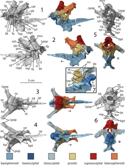
In non-human vertebrates, the foramen magnum is typically oriented toward the back rather than downward. These braincases often contain a higher number of endochondral bones rather than dermal bones. Key components include:
- Basioccipital: The rear lower part of the braincase. In reptiles and birds, it forms the occipital condyle, a ball-and-socket joint that connects to the vertebrae.
- Exoccipitals: Paired bones at the rear. In mammals and amphibians, these have evolved into paired occipital condyles.
- Supraoccipital: The rear upper part, homologous to the squamous part of the human occipital bone.
- Opisthotics: Paired bones forming the rear lateral part, often fusing with the exoccipital near the jugular foramen.
- Prootics: Lateral bones that house the exit for the trigeminal nerve (V). These are homologous to the petrous part of the human temporal bone.
- Basisphenoid: The front lower part, featuring basipterygoid processes that link to the bony palate.
- Parasphenoid: A flat dermal plate that often fuses with the basisphenoid to form the parabasisphenoid.
| Bone Name | Quantity | Location/Role |
|---|---|---|
| Frontal | 1 | Forehead/Upper front |
| Parietal | 2 | Upper sides and roof |
| Temporal | 2 | Lower sides/Ear region |
| Occipital | 1 | Back and base |
| Sphenoid | 1 | Central base (butterfly-shaped) |
| Ethmoid | 1 | Between the eyes/Nasal cavity |
Frequently Asked Questions
What is the difference between the neurocranium and the facial skeleton?
The neurocranium is the upper and back part of the skull that protects the brain, whereas the facial skeleton (splanchnocranium) forms the structure of the face.
How many bones make up the human neurocranium?
The human neurocranium is typically composed of eight bones: one frontal, two parietal, two temporal, one occipital, one sphenoid, and one ethmoid.
What is the chondrocranium?
The chondrocranium is the cartilaginous part of the neurocranium that forms the bones at the base of the skull.
How does the neurocranium differ between humans and other vertebrates?
In humans, the neurocranium has expanded to cover the upper part of the skull to accommodate a larger brain, and the foramen magnum is oriented downward. In other vertebrates, the foramen magnum usually faces backward, and the braincase often contains more endochondral bones.
What is the function of the foramen magnum?
The foramen magnum is a large opening at the base of the neurocranium that allows the neural spine (spinal cord) to connect to the brain.