Neural Tube Development and Patterning in Vertebrates
The neural tube is the fundamental embryonic precursor to the central nervous system (CNS), which eventually forms the brain and spinal cord. In chordates, including humans, the process begins as a neural groove that deepens and closes to form a hollow tube. In humans, this critical closure typically occurs by the fourth week of pregnancy, specifically around the 28th day after conception.
Key Facts
- Function: Serves as the embryonic foundation for the brain and spinal cord.
- Timing: Closes by the 28th day after conception in humans.
- Primary Neurulation: Involves the folding of the neural plate into a tube.
- Secondary Neurulation: Involves the formation of a solid cord that later hollows out.
- Key Signaling Molecules: Sonic hedgehog (Shh) patterns the ventral side, while Bone Morphogenic Proteins (BMPs) pattern the dorsal side.
- Clinical Significance: Failure of the tube to close leads to neural tube defects (NTDs) like spina bifida and anencephaly.
The Process of Neurulation
Neurulation, the process of forming the neural tube, occurs via two distinct mechanisms: primary and secondary neurulation. The extent to which each is used varies by species.
Primary Neurulation
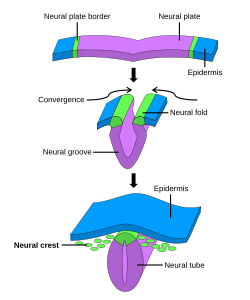
Primary neurulation begins with the formation of the neural plate. The edges of this plate thicken and lift to create neural folds, while the center remains grounded, forming a U-shaped neural groove. As the folds pinch toward the midline and fuse, they divide the ectoderm into three distinct cell types:
- Neural Tube: The internal structure that becomes the CNS.
- Epidermis: The external skin layer.
- Neural Crest Cells: Cells located between the tube and epidermis that migrate to various locations in the body.
Secondary Neurulation
In secondary neurulation, cells form a solid, cord-like structure that migrates inside the embryo and subsequently hollows out to create the tube. While fish rely solely on this method, mammals use it for the posterior regions of the tube, typically beginning around the 35th somite. In avian species, the anterior regions develop via primary neurulation, while posterior regions use secondary neurulation.
Structural Evolution of the CNS
As the neural tube develops, neuroepithelial cells divide to create four primary subdivisions that evolve into the adult central nervous system.
- Prosencephalon (Forebrain): Further divides into the telencephalon (cerebrum) and diencephalon (hypothalamus and optic vesicles).
- Mesencephalon (Midbrain): Remains as the midbrain.
- Rhombencephalon (Hindbrain): Develops into the metencephalon (pons and cerebellum) and the myelencephalon (medulla oblongata).
- Spinal Cord: Develops from the posterior portion of the tube.
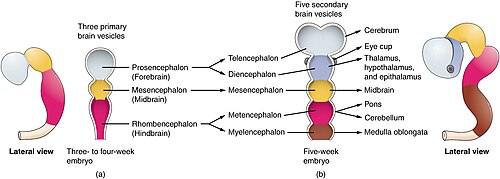
Initially, the tube remains open at both the cranial and caudal ends via openings called neuropores. These must close during the fourth week of human development; failure to do so results in neural tube defects (NTDs).
Functional Zoning: Alar and Basal Plates
The tube is organized functionally along its diameter. The alar plate (dorsal part) is primarily associated with sensory functions, while the basal plate (ventral part) is associated with motor control.
Dorsal-Ventral Patterning
The neural tube uses a complex system of signaling molecules to tell progenitor cells what type of neuron to become. This is often described by the French flag model of morphogenesis, where concentration gradients determine cell fate.
Ventral Patterning and Sonic Hedgehog (Shh)
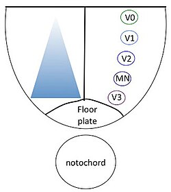
Sonic hedgehog (Shh) is a morphogen—a molecule that acts in a concentration-dependent manner. Secreted first by the notochord and later by the floor plate cells, Shh creates a gradient that specifies ventral cell types, including floor plate cells, motor neurons, and various interneurons (V0-V3).
Shh regulates transcription factors in two classes: Class I proteins are inhibited by Shh, while Class II proteins are activated. The interaction between these classes defines the boundaries of neuronal identity. Additionally, retinoic acid works with Shh to induce Pax6 and Olig2 during motor neuron differentiation.
Dorsal Patterning and BMPs
The dorsal axis is patterned primarily by Bone Morphogenic Proteins (BMPs) and Wnt family members. BMPs are initially secreted by the overlying ectoderm and later by the roof plate. Similar to Shh, BMPs act in a concentration-dependent manner; for example, a deficiency in BMP signaling in zebrafish leads to a loss of dorsal sensory neurons and an increase in interneurons.
Immune System Interactions
Recent research indicates that the immune system plays a role during neural tube development. Macrophages, which exhibit both pro-inflammatory and anti-inflammatory functions, interact with developing neural cells via cytokine signaling and pathways such as NF-κB and JAK–STAT. Alterations in these immune interactions may contribute to the development of NTDs, making certain immune-regulation genes potential prenatal diagnostic markers.
| Feature | Ventral Side | Dorsal Side |
|---|---|---|
| Primary Plate | Basal Plate | Alar Plate |
| Primary Function | Motor Control | Sensation |
| Key Signaling Molecule | Sonic hedgehog (Shh) | BMPs / Wnt |
| Key Structures | Floor plate, Motor neurons | Roof plate, Sensory neurons |
Frequently Asked Questions
What is the difference between primary and secondary neurulation?
Primary neurulation involves the folding of a neural plate into a tube, whereas secondary neurulation involves the formation of a solid cord of cells that subsequently hollows out to form the tube.
What happens if the neuropores do not close?
If the neuropores (the openings at the ends of the neural tube) fail to close during the fourth week of development, it can result in neural tube defects such as anencephaly or spina bifida.
How does Sonic hedgehog (Shh) determine cell type?
Shh acts as a morphogen, meaning it creates a concentration gradient. Depending on the concentration of Shh a cell is exposed to and the duration of that exposure, the cell will activate or inhibit specific transcription factors to become a specific type of ventral neuron.
Which parts of the brain develop from the rhombencephalon?
The rhombencephalon, or hindbrain, develops into the metencephalon (which becomes the pons and cerebellum) and the myelencephalon (which becomes the medulla oblongata).
Do all animals use the same neurulation process?
No. For example, fish proceed only via secondary neurulation, while mammals and birds use a combination of both primary and secondary neurulation depending on the region of the tube.