Nervous System: Evolution, Structure, and Biological Function
The nervous system is a complex network of specialized cells and organs that coordinate an organism's actions and sensory perceptions. From the simplest nerve nets in primitive animals to the highly sophisticated human brain, this system allows living beings to interact with their environment, process information, and maintain internal homeostasis.
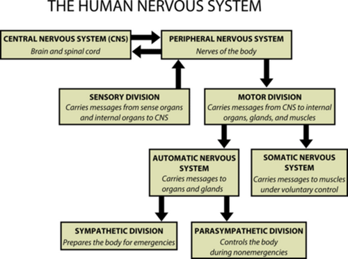
At its core, the nervous system relies on the transmission of electrochemical signals. In humans, this involves a sophisticated division between the central nervous system (the brain and spinal cord) and the peripheral nervous system, which connects the center to the rest of the body.
Key Facts
- Bilaterians share a common ancestor from the Ediacaran period (550–600 million years ago) with a basic nerve cord and brain structure.
- The nematode C. elegans has a fully mapped connectome, with exactly 302 neurons in hermaphrodites and 383 in males.
- Neural induction is driven by the inhibition of bone morphogenetic protein (BMP), specifically BMP4, by proteins like Noggin and Chordin.
- Neurotrophins are signaling molecules found in both vertebrates and invertebrates that regulate neuron growth and survival.
- Synaptic transmission occurs when an action potential triggers the release of neurotransmitters across a synapse to a target cell.
Comparative Anatomy and Evolution

The Bilaterian Blueprint
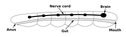
Most existing animals are bilaterians—organisms with left and right sides that are approximate mirror images. Evidence suggests they descended from a wormlike ancestor appearing 550–600 million years ago. The fundamental body plan consists of a hollow gut cavity and a nerve cord featuring segmental enlargements called ganglia (clusters of nerve cell bodies), with a primary ganglion at the front serving as the brain.
Diversity Across Species
The complexity of nervous systems varies wildly across the animal kingdom. In the roundworm Caenorhabditis elegans, the entire neural network is known, revealing sexual dimorphism where males possess more neurons than hermaphrodites to perform sex-specific functions.
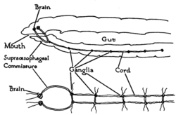
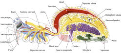
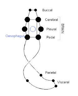
Other invertebrates, such as arthropods and molluscs, exhibit distinct neural architectures. Spiders possess a centralized system, while molluscs utilize a more simplified arrangement of ganglia to manage their bodily functions.
Human Neural Architecture
Cellular Components
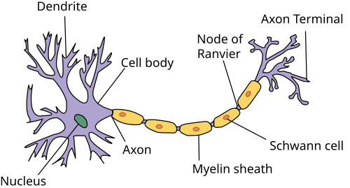
The human nervous system is composed primarily of neurons (cells that transmit electrical impulses) and glial cells (support cells that maintain homeostasis and protect neurons). Neurons are structured to send signals over long distances via axons, often insulated by Schwann cells in the peripheral nervous system to increase signal speed.
Protective Structures and Organization
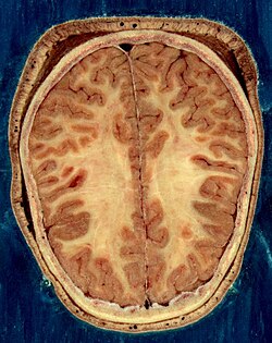
The brain and spinal cord are highly vulnerable and are therefore encased in bone (the skull and vertebrae) and protected by multiple layers of membranes and fluid.
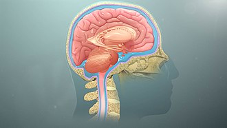
The brain's composition includes gray matter, consisting mainly of neuronal cell bodies, and white matter, which consists of myelinated axons that connect different brain regions.
Biological Function and Mechanisms
Synaptic Transmission
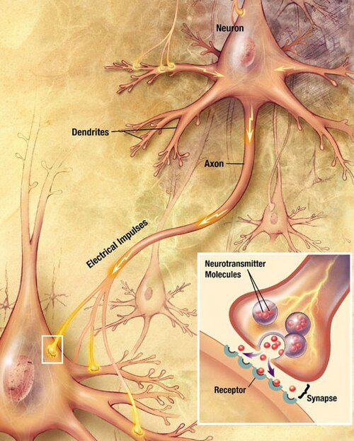
Communication between neurons occurs at the synapse. An electrochemical wave, known as an action potential, travels along the axon. Upon reaching the synapse, it triggers the release of neurotransmitter molecules, which bind to receptors on the target cell's membrane, continuing the signal.
Sensory and Motor Pathways
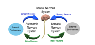
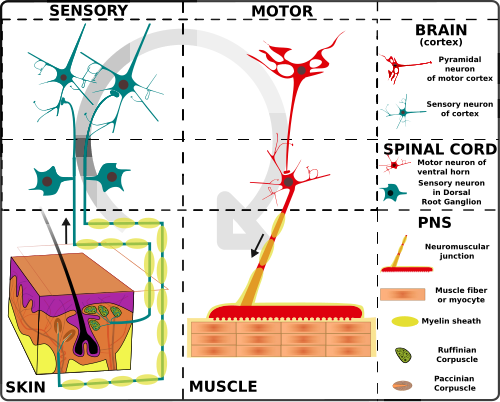
The system operates in a loop: sensory receptors pick up external stimuli and send signals to the spinal cord and brain. After processing, the brain sends signals back through the spinal cord to motor neurons, which trigger a physical response.
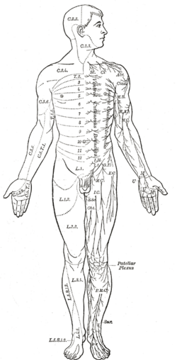
Specific areas of the body are innervated by specific spinal nerves, creating a map of sensory and motor control across the human surface.
Advanced Neural Circuits
Beyond basic reflexes, the brain utilizes mirror neurons—specialized cells that fire both when an individual performs an action and when they observe someone else performing that same action. These are thought to play a role in imitation and understanding the intentions of others.
Development and Neural Induction
The formation of nervous tissue is a process called neural induction. Research indicates that this occurs when the organizer region (a group of mesodermal cells) inhibits the gene for bone morphogenetic protein (BMP), specifically BMP4. Proteins such as Noggin and Chordin facilitate this inhibition, allowing the ectoderm to transform into neural tissue.
Furthermore, a family of molecules called neurotrophins (such as DNT1 in flies) regulates the survival and growth of neurons. Because these are found in both vertebrates and invertebrates, they likely represent an ancient mechanism for nervous system formation common to all bilaterians.
| Component/Process | Description | Key Molecule/Cell |
|---|---|---|
| Neural Induction | Process of forming nervous tissue from ectoderm | Noggin, Chordin, BMP4 |
| Signal Transmission | Electrochemical communication between neurons | Action Potential, Neurotransmitters |
| Neuron Survival | Regulation of growth and survival of nerve cells | Neurotrophins (e.g., DNT1) |
| Structural Support | Maintenance and protection of neurons | Glial Cells, Schwann Cells |
Frequently Asked Questions
What is the difference between gray matter and white matter?
Gray matter consists primarily of neuronal cell bodies and dendrites, where processing occurs. White matter consists of myelinated axons, which act as the "cables" that transmit signals between different areas of gray matter.
How do mirror neurons work?
Mirror neurons are a class of cells that activate both when an organism performs a specific action and when it observes another organism performing that same action, potentially aiding in learning and empathy.
What role does BMP4 play in the development of the nervous system?
BMP4 (bone morphogenetic protein 4) typically inhibits the formation of neural tissue. For the nervous system to develop, proteins like Noggin and Chordin must inhibit BMP4, allowing the ectoderm to become neural tissue.
What is a connectome?
A connectome is a comprehensive map of all the neural connections (synapses) within a nervous system. A complete connectome has been mapped for the roundworm C. elegans.
How is a signal transmitted from one neuron to another?
A signal travels as an electrical action potential down the axon. When it reaches the synapse, it triggers the release of chemical neurotransmitters that cross the gap and bind to receptors on the next cell, converting the electrical signal back into a chemical one and then potentially back to electrical.