Muscle Tissue: Types, Structure, and Biological Functions
Muscle is a specialized soft tissue and one of the four basic types of animal tissues. Its primary biological role is to facilitate movement through the ability to contract and relax. This process is driven by the interaction of contractile proteins—specifically actin and myosin—along with regulatory proteins known as troponin and tropomyosin. The formation of these tissues during embryonic development is a process called myogenesis.
The etymology of the word "muscle" derives from the Latin musculus, a diminutive of mus (mouse), because a flexed biceps muscle resembles the back of a mouse. This same linguistic root is found in the Greek word mȳs.
Key Facts
- Three Vertebrate Types: Skeletal, cardiac, and smooth muscle.
- Control Mechanisms: Skeletal muscle is voluntary; cardiac and smooth muscles are involuntary.
- Striation: Skeletal and cardiac muscles are striated (striped), while smooth muscle is non-striated.
- Composition: Skeletal muscle makes up approximately 42% of an average adult man's body mass and 36% of an average adult woman's.
- Density: Muscle tissue is about 15% denser than adipose (fat) tissue.
Types of Muscle Tissue in Vertebrates
Skeletal Muscle
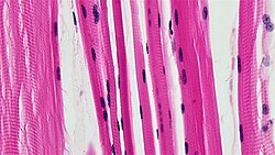
Skeletal muscle is a striated, voluntary tissue consisting of elongated, multinucleate cells called muscle fibers. These fibers are arranged in parallel bundles of myofibrils containing sarcomeres—the basic contractile units that create the tissue's striped appearance. Skeletal muscles are anchored to bones via tendons or aponeuroses to enable locomotion and maintain posture.
While most skeletal muscle movement is conscious, reflexes are a form of non-conscious activation that still occurs via the central nervous system.
Cardiac Muscle
Found exclusively in the heart walls as the myocardium, cardiac muscle is striated but involuntary. Unlike the parallel arrangement of skeletal muscle, cardiac cells connect at irregular, branching angles called intercalated discs. This tissue is regulated by the autonomic nervous system and relies on the coronary arteries for a constant supply of oxygen and nutrients to maintain rhythmic contractions (systole).
Smooth Muscle
Smooth muscle is non-striated and involuntary. It is located in the walls of hollow organs and tubular structures, including the stomach, intestines, bladder, uterus, and blood vessels. It also controls the arrector pili in the skin, which causes body hair to stand up. Smooth muscle is divided into single-unit (contracting as a syncytium) and multiunit (allowing for fine, individual cell control) types.
Comparative Analysis of Muscle Tissues
| Feature | Smooth Muscle | Cardiac Muscle | Skeletal Muscle |
|---|---|---|---|
| Striation | None | Present | Present |
| Control | Involuntary | Involuntary | Voluntary |
| Fiber Shape | Fusiform, short | Branching | Cylindrical, long |
| Nuclei | 1 | Usually 1 (some 2-4) | Many (>1) |
| Sarcomeres | None | Present | Present |
| ATPase Level | Little | Moderate | Abundant |
Skeletal Muscle Fiber Classifications
Skeletal muscles are categorized into two primary fiber types based on their metabolic and contractile properties:
- Type I (Slow-twitch): Also known as red muscle, these are rich in myoglobin, mitochondria, and capillaries. They are designed for aerobic activity and endurance.
- Type II (Fast-twitch): These provide rapid, powerful contractions. They are further divided into:
- Type IIa: Aerobic and rich in mitochondria (red).
- Type IIx: The fastest human muscle type; capable of short, anaerobic bursts of force.
- Type IIb: Anaerobic "white" muscle, predominant in small animals like rodents.
Embryonic Development
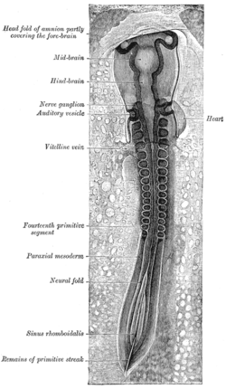
All muscles originate from the paraxial mesoderm, which divides into segments called somites. Each somite splits into three parts: the sclerotome (vertebrae), dermatome (skin), and myotome (muscle). The myotome further divides into the epimere (forming epaxial muscles like the erector spinae) and the hypomere (forming hypaxial muscles, including the limbs).
Muscle progenitor cells, called myoblasts, either stay in the somite or migrate to specific locations guided by chemical signals, where they fuse to form elongated skeletal muscle cells.
Invertebrate Muscle Variations
Invertebrates possess three types of muscle based on striation: transversely striated, obliquely striated, and smooth. Notably, arthropods lack smooth muscle entirely. The transversely striated type is the most similar to vertebrate skeletal muscle.
Frequently Asked Questions
What is the difference between voluntary and involuntary muscles?
Voluntary muscles, such as skeletal muscle, are controlled consciously by the central nervous system. Involuntary muscles, such as cardiac and smooth muscle, operate without conscious effort, regulated instead by the autonomic nervous system, hormones, or internal pacemakers.
What are sarcomeres and why are they important?
Sarcomeres are the basic contractile units of striated muscle. Their regular, repeating arrangement gives skeletal and cardiac muscle their striped appearance and allows the muscle to shorten during contraction.
Why is Type I muscle red and Type IIb muscle white?
Type I fibers are red because they contain high levels of myoglobin and mitochondria to support aerobic metabolism. Type IIb fibers are white because they have lower concentrations of these oxygen-binding proteins and rely on anaerobic glycolysis.
Where is smooth muscle located in the human body?
Smooth muscle is found in the walls of hollow organs (stomach, bladder, uterus), tubular structures (blood vessels, lymph vessels, bile ducts), and the iris of the eye.
How does muscle density compare to fat?
Muscle tissue has a density of approximately 1.06 kg/liter, while adipose tissue is about 0.9196 kg/liter, making muscle roughly 15% denser than fat.