Coelom: The Biological Role and Evolution of Animal Body Cavities
In the diverse world of zoology, the structural organization of an animal's interior is a fundamental aspect of its biology. One of the most critical features is the coelom (pronounced SEE-ləm), the primary body cavity found in many animals. Positioned inside the body, the coelom surrounds and contains the digestive tract and other vital organs, providing a protected environment for biological processes to occur.
Derived from the Ancient Greek word koilía, meaning 'cavity,' the coelom is more than just an empty space. In many species, it is lined with mesothelium (a specialized membrane), while in others, such as molluscs, it remains undifferentiated. This structural feature has historically served as a primary tool for biologists to categorize bilaterian animal phyla.
Key Facts
- Definition: A fluid-filled body cavity lined with tissue derived from the mesoderm.
- Primary Function: Acts as a hydrostatic skeleton, absorbs shock, and allows internal organs to grow and move independently of the body wall.
- Development: Forms via schizocoely in protostomes and enterocoely in deuterostomes.
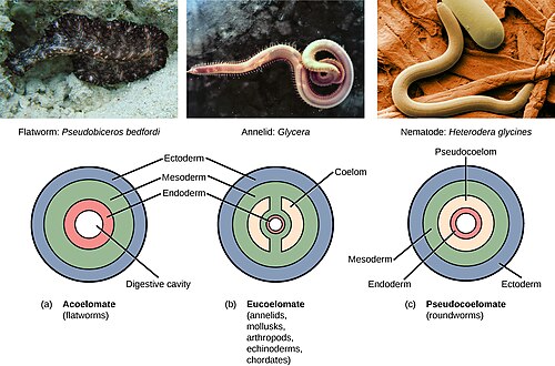
- Classification: Animals are broadly described as coelomates (true cavity), pseudocoelomates (partial lining), or acoelomates (no cavity).
- Fluid Role: Coelomic fluid transports nutrients, gases, and waste, and stores gametes during maturation.
Development and Formation
The coelom is defined as the mesodermally lined cavity situated between the gut and the outer body wall. Its formation begins during the gastrulation stage of embryonic development, starting with the creation of the archenteron, a blind pouch that becomes the developing digestive tube.
Protostomes and Schizocoely
In protostomes, the coelom forms through a process called schizocoely. During this process, the mesoderm splits into two distinct layers. The first layer attaches to the body wall or ectoderm, forming the parietal layer, while the second surrounds the endoderm (the alimentary canal), forming the visceral layer. The resulting space between these two layers is the coelom.
Deuterostomes and Enterocoely
Deuterostomes utilize a different method known as enterocoely. In these organisms, the wall of the archenteron produces buds of mesoderm. These mesodermal diverticula then hollow out to create the coelomic cavities. Animals that develop this way are called enterocoelomates. Major examples include chordates (vertebrates, tunicates, and lancelets), echinoderms (starfish and sea urchins), and hemichordates (acorn worms).
Functions of the Coelom
The presence of a coelom provides several evolutionary advantages that allow animals to grow larger and more complex:
- Physical Support: It can provide a hydrostatic skeleton, using fluid pressure to maintain shape and facilitate movement.
- Organ Independence: It allows muscles to grow independently of the body wall. For example, in tardigrades (water bears), the digestive tract is suspended within the body by the mesentery.
- Protection: The cavity acts as a shock absorber for internal organs.
- Immune Support: It supports an immune system via coelomocytes, which are specialized cells that either float freely or attach to the cavity wall.
The Role of Coelomic Fluid
The fluid filling the cavity, known as coelomic fluid, is circulated by mesothelial cilia or the contraction of body wall muscles. This fluid is essential for the transport of nutrients, gases, and waste products. Additionally, it serves as a reservoir for waste and a storage area for sperm and eggs during their maturation process.
Zoological Classification
While modern molecular phylogeny has moved away from using body cavities as a formal taxonomic system, the terms remain useful for describing animal morphology. Animals are generally categorized into three informal groups:
| Group | Cavity Type | Lining Description | Example Phyla |
|---|---|---|---|
| Coelomata | True Coelom | Completely lined by mesoderm (peritoneum) | Chordata, Annelida, Arthropoda |
| Pseudocoelomata | Pseudocoelom | Partially lined by mesoderm | Nematoda, Rotifera |
| Acoelomata | None | No cavity; filled with semi-solid mesoderm | Platyhelminthes (Flatworms) |
Coelomates (Eucoelomates)
These animals possess a true coelom with a complete lining called the peritoneum. This allows organs to be suspended in a specific order while remaining free to move. Most bilateral animals, including all vertebrates, fall into this category. Subtypes include the schizocoelom (found in annelids and molluscs), the haemocoelom (a blood-filled cavity in arthropods), and the enterocoelom (found in chordates).
Pseudocoelomates (Blastocoelomates)
Pseudocoelomates have a fluid-filled cavity that is only partially lined with mesoderm. Because they lack a vascular blood system, they rely on diffusion and osmosis to circulate nutrients. They typically lack a skeleton, relying instead on hydrostatic pressure for support. Examples include roundworms (Nematoda) and horsehair worms (Nematomorpha).
Acoelomates
Acoelomates, such as flatworms, have no body cavity. Their organs are held in place by semi-solid mesodermal tissues. While this leaves their organs vulnerable to crushing forces, their dorso-ventral flattening creates a high surface-area-to-volume ratio, allowing them to exchange gases and nutrients through simple diffusion.
Frequently Asked Questions
What is the difference between a coelom and a pseudocoelom?
A true coelom is completely lined with tissue derived from the mesoderm, allowing for complex organ suspension and movement. A pseudocoelom is only partially lined with mesoderm, resulting in a less organized internal structure.
How does a hydrostatic skeleton work?
A hydrostatic skeleton uses the incompressible nature of the coelomic fluid. By contracting muscles against this fluid-filled cavity, the animal can create structural rigidity and generate movement.
Which animals are considered acoelomates?
Flatworms (Platyhelminthes) are the primary example of acoelomates. Other examples include Micrognathozoa, Mesozoa, and Xenacoelomorpha.
What are the two main ways a coelom develops in embryos?
The two methods are schizocoely, where the mesoderm splits to form the cavity (common in protostomes), and enterocoely, where the cavity forms from buds of the embryonic gut (common in deuterostomes).
Why is the coelom important for larger animals?
The coelom allows for the development of larger, more complex organs that can grow and function independently of the outer body wall, and it provides a means of transporting nutrients and waste in animals too large to rely solely on diffusion.