Animal Brains: Evolution, Structure, and Function Across Species

Animal Brains: Evolution, Structure, and Function Across Species

The brain is the most complex organ in the animal kingdom, serving as the command center for the nervous system. From the simple nerve cords of early bilaterians to the highly folded neocortex of primates, the brain has evolved to process sensory information, coordinate movement, and enable complex behaviors. By studying the diversity of brain structures across different species, scientists can uncover the fundamental principles of how life perceives and interacts with the world.

Key Facts
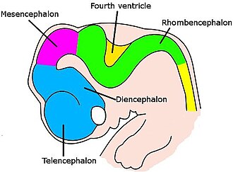
Very simple drawing of the front end of a human embryo, showing each vesicle of the developing brain in a different color.
Brain of a human embryo in the sixth week of development
  • Encephalization Quotient (EQ) is used to compare relative brain size across species; humans have the highest EQ (7.4–7.8).
  • Neurotransmitters are chemical messengers that cross synapses to alter the electrical activity of target cells.
  • Vertebrate brains share a common embryonic origin, differentiating into the forebrain, midbrain, and hindbrain.
  • Neural plasticity allows the brain to change its structure and function in response to environmental enrichment.
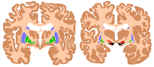
  • The Basal Ganglia and Cerebellum are critical for action selection and movement precision, respectively.

Cellular Structure and Physiology
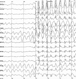
Graph showing 16 voltage traces going across the page from left to right, each showing a different signal. At the middle of the page all of the traces abruptly begin to show sharp jerky spikes, which continue to the end of the plot.
Brain electrical activity recorded from a human patient during an epileptic seizure

At its core, the brain is composed of neurons that generate electrical signals. These signals travel along axons to reach a synapse—a junction where neurotransmitters are released to communicate with other cells.
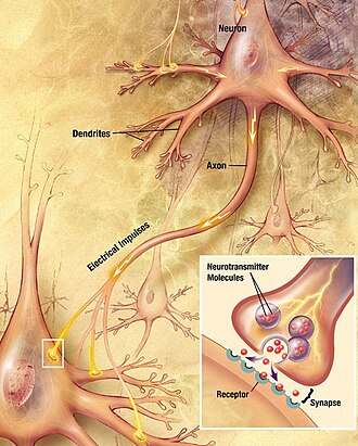
drawing showing a neuron with a fiber emanating from it labeled "axon" and making contact with another cell. An inset shows an enlargement of the contact zone.
Neurons generate electrical signals that travel along their axons. When an electrical impulse reaches a junction called a synapse, it causes a neurotransmitter to be released, which binds to receptors on other cells and thereby alters their electrical activity.

The interaction between these neurons is regulated by receptors. For example, GABA receptors play a vital role in inhibitory signaling, helping to balance the brain's electrical activity.
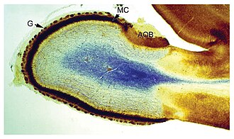
a blob with a blue patch in the center, surrounded by a white area, surrounded by a thin strip of dark-colored material
Cross section of the olfactory bulb of a rat, stained in two different ways at the same time: one stain shows neuronal cell bodies, the other shows receptors for the neurotransmitter GABA.

Beyond electrical impulses, the brain requires significant metabolic support. While glucose is the primary fuel, some species and conditions utilize alternative fuels such as acetate, heptanoate, and octanoate to maintain neural function.

Evolution of the Brain

Gulai otak, beef brain curry from Indonesia

The Bilaterian Foundation

The most basic blueprint for a brain is found in bilaterian animals, characterized by a nerve cord with segmental enlargements and a concentrated group of neurons at the front of the body.
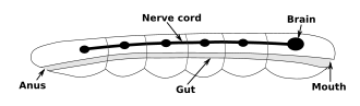
A rod-shaped body contains a digestive system running from the mouth at one end to the anus at the other. Alongside the digestive system is a nerve cord with a brain at the end, near to the mouth.
Nervous system of a generic bilaterian animal, in the form of a nerve cord with segmental enlargements, and a "brain" at the front

Invertebrate Complexity

Invertebrates exhibit a wide range of neural sophistication. Fruit flies (Drosophila) are frequently used in research to understand how genes drive brain development and structural plasticity.
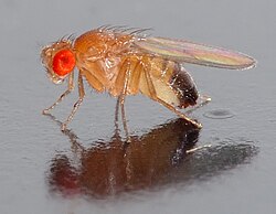
A fly resting on a reflective surface. A large, red eye faces the camera. The body appears transparent, apart from black pigment at the end of its abdomen.
Fruit flies (Drosophila) have been extensively studied to gain insight into the role of genes in brain development.

Vertebrate Diversification

Vertebrate brains develop from a set of embryonic subdivisions that later differentiate into adult structures. While the basic parts are conserved across species, their size and shape vary drastically based on the animal's ecological needs.
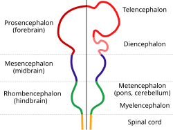
The nervous system is shown as a rod with protrusions along its length. The spinal cord at the bottom connects to the hindbrain which widens out before narrowing again. This is connected to the midbrain, which again bulges, and which finally connects to the forebrain which has two large protrusions.
The main subdivisions of the embryonic vertebrate brain (left), which later differentiate into structures of the adult brain (right)

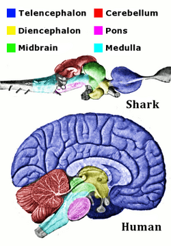
For instance, sharks possess a brain structure that shares the same fundamental regions as humans, though the proportions differ significantly.
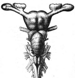
A T-shaped object is made up of the cord at the bottom which feeds into a lower central mass. This is topped by a larger central mass with an arm extending from either side.
The brain of a shark
Corresponding regions of human and shark brain are shown. The shark brain is splayed out, while the human brain is more compact. The shark brain starts with the medulla, which is surrounded by various structures, and ends with the telencephalon. The cross-section of the human brain shows the medulla at the bottom surrounded by the same structures, with the telencephalon thickly coating the top of the brain.
The main anatomical regions of the vertebrate brain, shown for shark and human. The same parts are present, but they differ greatly in size and shape.

Comparing reptiles and birds reveals further specialization. Birds often have highly developed visual processing areas to support flight and navigation.
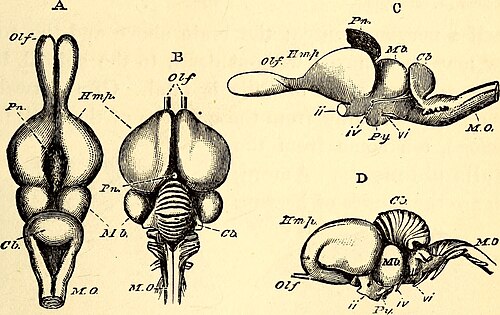
Anatomical comparison between the brain of a lizard (A and C) and the brain of a turkey (B and D). Abbreviations: Olf, olfactory lobes; Hmp, cerebral hemispheres; Pn, pineal gland; Mb, optic lobes of the middle brain; Cb, cerebellum; MO, medulla oblongata; ii, optic nerves; iv and vi, nerves for the muscles of the eye; Py, pituitary body.
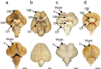
Brains of an emu, a kiwi, a barn owl, and a pigeon, with visual processing areas labelled

Mammalian and Primate Brains

Mammals, particularly primates, show a significant increase in the complexity of the forebrain. The Encephalization Quotient (EQ) provides a metric for this relative brain size compared to body mass.

Relative Brain Size (EQ) Across Selected Species
Species Encephalization Quotient (EQ)
Human 7.4–7.8
Bottlenose dolphin 4.14
Common chimpanzee 2.2–2.5
Elephant 1.13–2.36
Dog 1.2
Rat 0.4

Brain Function and Motor Control

The brain is organized into specialized regions that handle different aspects of survival and interaction. Motor control, for example, is a distributed process involving several key areas:

  • Ventral horn (Spinal cord): Directly activates muscles.
  • Cerebellum: Calibrates the timing and precision of movements.
  • Basal ganglia: Selects actions based on motivation.
  • Motor cortex: Activates spinal motor circuits.
  • Prefrontal cortex: Manages planning and executive functions.
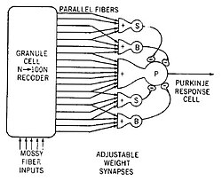
Model of a neural circuit in the cerebellum, as proposed by James S. Albus
Components of the basal ganglia, shown in two cross-sections of the human brain. Blue: caudate nucleus and putamen. Green: globus pallidus. Red: subthalamic nucleus. Black: substantia nigra.

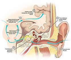
Other critical regions include the hypothalamus, which manages homeostasis, and the auditory system, which processes sound signals through complex neural circuits.
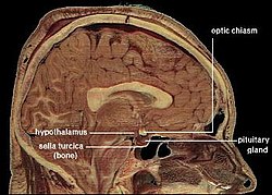
Cross-section of a human head, showing location of the hypothalamus
Drawing showing the ear, inner ear, and brain areas involved in hearing. A series of light blue arrows shows the flow of signals through the system.
Diagram of signal processing in the auditory system

Modern Research and History

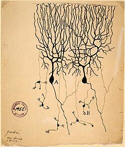
Our understanding of the brain has evolved from the early anatomical drawings of Andreas Vesalius in 1543 to the detailed cellular sketches of Santiago Ramón y Cajal.
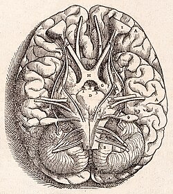
Andreas Vesalius' Fabrica, published in 1543, showing the base of the human brain, including optic chiasma, cerebellum, olfactory bulbs, etc.
A drawing on yellowing paper with an archiving stamp in the corner. A spidery tree branch structure connects to the top of a mass. A few narrow processes follow away from the bottom of the mass.
Drawing by Santiago Ramón y Cajal of two types of Golgi-stained neurons from the cerebellum of a pigeon

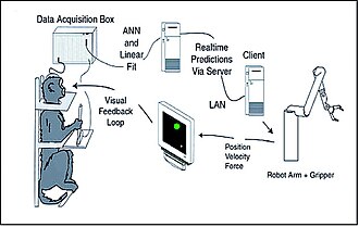
Today, neuroscience employs advanced technology to map the brain. The Human Brain Project, launched in 2013, aims to simulate the entire human brain, while other researchers develop brain-computer interfaces that allow animals to control robotic limbs using neural activity.
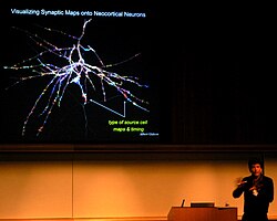
The Human Brain Project is a large scientific research project, starting in 2013, which aims to simulate the complete human brain.
Drawing showing a monkey in a restraint chair, a computer monitor, a rototic arm, and three pieces of computer equipment, with arrows between them to show the flow of information.
Design of an experiment in which brain activity from a monkey was used to control a robotic arm[135]

Frequently Asked Questions

What is the Encephalization Quotient (EQ)?

The EQ is a measure of relative brain size, calculated by comparing the actual brain mass of a species to the expected brain mass for an animal of that body size. A higher EQ generally suggests a higher proportion of neurons dedicated to complex cognitive functions.

How do neurons communicate with each other?

Neurons communicate via electrical impulses that travel along axons. When the impulse reaches a synapse, it triggers the release of neurotransmitters, which bind to receptors on the receiving cell to either excite or inhibit its activity.

What is the role of the cerebellum in movement?

The cerebellum is primarily responsible for the calibration of movement. It ensures that motor actions are precise and timed correctly, allowing for smooth coordination.

Do all vertebrates have the same basic brain structure?

Yes, all vertebrates share the same fundamental embryonic subdivisions that develop into the forebrain, midbrain, and hindbrain. However, these regions vary greatly in size and shape depending on the species.

What is the purpose of the prefrontal cortex?

The prefrontal cortex is the center for higher-order cognitive processes, including planning, decision-making, and other executive functions.